Review
. 2025 May 12;32(1):48.
doi: 10.1186/s12929-025-01142-w.

Decoding innate lymphoid cells and innate-like lymphocytes in asthma: pathways to mechanisms and therapies

Affiliations
Review

Decoding innate lymphoid cells and innate-like lymphocytes in asthma: pathways to mechanisms and therapies

Christina Li-Ping Thio et al. J Biomed Sci. .

Abstract

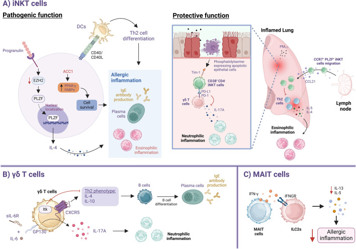
Asthma is a chronic inflammatory lung disease driven by a complex interplay between innate and adaptive immune components. Among these, innate lymphoid cells (ILCs) and innate-like lymphocytes have emerged as crucial players in shaping the disease phenotype. Within the ILC family, group 2 ILCs (ILC2s), in particular, contribute significantly to type 2 inflammation through their rapid production of cytokines such as IL-5 and IL-13, promoting airway eosinophilia and airway hyperreactivity. On the other hand, innate-like lymphocytes such as invariant natural killer T (iNKT) cells can play either pathogenic or protective roles in asthma, depending on the stimuli and lung microenvironment. Regulatory mechanisms, including cytokine signaling, metabolic and dietary cues, and interactions with other immune cells, play critical roles in modulating their functions. In this review, we highlight current findings on the role of ILCs and innate-like lymphocytes in asthma development and pathogenesis. We also examine the underlying mechanisms regulating their function and their interplay with other immune cells. Finally, we explore current therapies targeting these cells and their effector cytokines for asthma management.

Keywords: Asthma; ILCs; Immune regulation; Innate-like lymphocytes; Therapeutic targets.


Conflict of interest statement

Declarations. Ethics approval and consent to participate: Not applicable. Consent for publication: Not applicable. Competing interests: The authors declare no competing interests.

Figures

Fig. 1
Fig. 1
Modulation of ILC function in asthma. A ILC2 activation is influenced by cytokines and other mediators. IL-33, TSLP, and IL-25 are primary activators, while IL-2, IL-7, IL-9, IL-4, and TNF superfamily members act as co-factors. Meanwhile, type I/II IFNs, IL-27, and IL-10 inhibit ILC2s. Activation leads to GATA3 and NF-κB phosphorylation, driving IL-13 and IL-5 production, proliferation, and survival. IL-1β, IL-12, and IL-18 promote ILC1-like differentiation, marked by IFN-γ production and T-bet expression, while IL-1β, IL-23, and TGF-β drive polarization into IL-17A-producting ILC3-like cells. Notably, IL-4 counteracts these effects. Non-cytokine regulators, including neuropeptides (NMU, VIP, CGRP), prostaglandins (PGI2, PGE2, PGD2), and leukotrienes, can either inhibit or enhance ILC2 activation. Butyrate inhibits ILC2s via HDAC suppression, while BHB indirectly suppresses ILC2s by limiting IL-2 from mast cells. Tregs (ICOS-ICOSL) and SLAMF receptors inhibit ILC2s, while Pla2g5+ macrophages (FFAs, IL-33) activate ILC2s. ILC2s also influence CD4+ T cells via BTN2A2, PD-L1, and OX40L, promoting Th2 differentiation. Notably, GATA3-expressing Tregs inhibit OX40L expression on ILC2s. B Resolvin-E1 promotes NK cell migration and enhances cytotoxic abilities, while lipoxin A4 promotes eosinophil apoptosis and clearance, together boosting pro-resolving functions of NK cells. In contrast, cannabinoids and PGI2 inhibit IFN-γ production by NK cells, subsequently attenuating ILC2 function. Type I IFNs from pDCs drive NK cell IFN-γ production. NK cell pathogenicity involves TLR3-induced IL-17A production, worsening asthma. NKG2D+ NK cells, via MULT-1, elevate IgE and eosinophil levels, promoting allergic airway inflammation. C ILC3s drive neutrophilic inflammation via IL-17. Cigarette smoke induces IL-1β from airway epithelium, generating memory-like ILC3s that worsen neutrophilic asthma. Conversely, MHC-II engagement on ILC3s inhibits microbe-specific Th17 and allergen-specific Th2 cells, reducing neutrophilic and eosinophilic asthma, respectively. NCR+ ILC3s interact with the microbiome to enhance protective functions via an unknown mechanism
Fig. 2
Fig. 2
Regulation of the pathogenic or protective functions of innate-like T cells in asthma. A IL-4 secretion by iNKT cells promotes allergic inflammation, with progranulin enhancing IL-4 production by downregulating EZH2 expression, thereby facilitating PLZF translocation and subsequent IL-4 expression. CD40-CD40L interaction between iNKT cells and DCs promotes Th2 differentiation, contributing to allergic inflammation. ACC1 mediates de novo fatty acid synthesis by upregulating PPARγ and FABPs, which promotes iNKT cell survival and enhances their deleterious functions. The protective functions of iNKT cells have been demonstrated in a PM2.5 model, where phosphatidylserine-expressing apoptotic epithelial cells activate suppressive CD38+ CD4 iNKT cells, leading to the upregulation of PD-L1 expression through interaction with Tim-1. PD-L1/PD-1 interaction between CD4 iNKT cells and γδ T cells inhibits IL-17A production by γδ T cells, thereby reducing neutrophilic inflammation. CCL21 can recruit CCR7+ PLZF+ iNKT cells into inflamed lungs to restrain Th2 response and mitigate eosinophilic inflammation. B CXCR5+ γδ T cells in allergic inflammation exhibit a Th2 phenotype (producing IL-4 and IL-10), promoting antibody production. Notably, Itk suppresses their development. IL-17A-producing γδ T cells contribute to neutrophilic inflammation via IL-6 trans-signaling, where IL-6 forms a complex with soluble IL-6R (sIL-6R) and binds to the GP130 receptor, amplifying inflammation. C MAIT cells produce IFN-γ which suppresses ILC2 cytokine production, attenuating airway inflammation

Similar articles

References

    1. Yang CH, Lv JJ, Li XY, Yang XT, Yin MY. Global burden of asthma in young adults in 204 countries and territories, 1990–2019: systematic analysis of the Global burden of disease study 2019. Prev Med Rep. 2024;37: 102531. - PMC - PubMed
    1. Davison J, Doe S. A patient case demonstrating the efficacy of benralizumab in uncontrolled severe eosinophilic asthma refractory to omalizumab and mepolizumab treatment. Respir Med Case Rep. 2021;34: 101557. - PMC - PubMed
    1. Lang DM. Severe asthma: epidemiology, burden of illness, and heterogeneity. Allergy Asthma Proc. 2015;36(6):418–24. - PubMed
    1. Kuruvilla ME, Lee FE, Lee GB. Understanding asthma phenotypes, endotypes, and mechanisms of disease. Clin Rev Allergy Immunol. 2019;56(2):219–33. - PMC - PubMed
    1. Ricciardolo FLM, Sprio AE, Baroso A, Gallo F, Riccardi E, Bertolini F, et al. Characterization of T2-low and T2-high asthma phenotypes in real-life. Biomedicines. 2021;9(11):1684. - PMC - PubMed