. 2025 Oct;77(10):1442-1450.
doi: 10.1002/art.43217. Epub 2025 Jun 27.

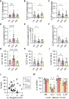
Innate Lymphoid Cell Phenotypic and Functional Alterations in Patients With Systemic Juvenile Idiopathic Arthritis

Affiliations

Innate Lymphoid Cell Phenotypic and Functional Alterations in Patients With Systemic Juvenile Idiopathic Arthritis

Linda Quatrini et al. Arthritis Rheumatol. 2025 Oct.

Abstract

Objective: Systemic juvenile idiopathic arthritis (sJIA) is a chronic childhood disease classically attributed to innate immune cell dysregulation. This study aimed to elucidate the role of innate lymphoid cells (ILCs), including natural killer (NK) cells and helper-ILCs (hILCs), in sJIA during clinically inactive disease (CID) through phenotypic and functional analysis.

Methods: Peripheral ILCs from children with sJIA during CID receiving interleukin-1 (IL-1) inhibitors (n = 40) were analyzed by flow cytometry and compared to 23 healthy children (HC) and 22 patients with unrelated autoinflammatory diseases taking IL-1 inhibitors. Plasma proteomic profiling was also performed.

Results: Patients with sJIA showed a significant reduction in circulating NK cell frequencies compared to HC, with an increased proportion of CD56bright NK cells. Although overall hILC frequencies were comparable to HC, ILC1s were increased, whereas ILC precursors were reduced. ILC1 frequency correlated positively with IL-18 plasma levels, whereas ILC2 frequency correlated negatively. Functional assessments revealed that NK cells from patients with sJIA had variable interferon γ (IFNγ) production upon IL-18/IL-12 stimulation, inversely correlating with IL-18 levels. Additionally, hILCs from these patients showed a specific impairment in IFNγ production despite normal IL-13 production, potentially linked to decreased IL-18 receptor α expression in ILC1s. Proteomic analysis confirmed IL-18 as the most up-regulated cytokine in sJIA plasma.

Conclusion: Patients with sJIA in CID exhibit significant innate immune abnormalities, including altered ILC subset distribution and impaired IFNγ production, strongly associated with IL-18 levels. These findings suggest ongoing immune dysregulation despite clinical remission, underscoring a potential role for ILCs and cytokine interaction in sJIA pathogenesis.


Figures

Figure 1
Figure 1
NK cell frequency and number in the peripheral blood of patients with sJIA are decreased compared to HC. (A–C) Scatter dot plots and bars showing the medians and interquartile ranges (IQR) of (A) NK cell frequency within live lymphocytes, (B) their absolute number, (C) and the frequency of CD56bright NK cells among total NK cells. D and E, Scatter dot plots and bars showing the medians and IQR of (D) hILC frequency within live lymphocytes and (E) their absolute number. Symbols represent individual patients. A, C, and D n = 23 HC; n = 31 sJIA; n = 14 AID. B and E n = 17 HC; n =26 sJIA; n =7 AID. Kruskal‐Wallis test was used, followed by uncorrected Dunn's test. (**P < 0.01; ***P < 0.005; ****P < 0.001). AID, autoinflammatory disease; HC, healthy children; hILC, helper innate lymphoid cell; NK, natural killer; sJIA, systemic juvenile idiopathic arthritis.
Figure 2
Figure 2
Changes in ILC subset frequencies are associated with IL‐18 plasma concentration. (A–I) Scatter dot plots and bars showing for each hILC subsets the medians and interquartile ranges (IQR) of (A–C) cell frequency within live lymphocytes, (D–F) of cell number, and (G–I) cell frequency within total hILCs. A–C and G–I n = 23 HC; n = 29 sJIA, n = 14 AID. D–F n = 17 HC; n = 26 sJIA, n = 7 AID. (L) Scatterplots showing the correlation between IL‐18 plasma concentration and ILC1 (empty dots) and ILC2 (black plots) frequency within hILCs. n = 29. r and P values were determined by the Spearman correlation test. Symbols represent individual patients. (M) Scatter dot plots and bars showing for each ILC subsets the median and IQR of IL‐18RA+ cell frequency within each ILC subset. n = 9 HC; n = 8 sJIA; n = 7 AID. In A–I and M, Kruskal‐Wallis test was used, followed by uncorrected Dunn's test (*P < 0.05; **P < 0.01; ***P < 0.005; ****P < 0.001). AID, autoinflammatory disease; HC, healthy children; hILC, helper innate lymphoid cell; ICL, innate lymphoid cell; IL‐18, interleukin 18; IL‐18RA, interleukin 18 receptor α; NK, natural killer; sJIA, systemic juvenile idiopathic arthritis.
Figure 3
Figure 3
Dysfunctional IFNγ production by ILCs in patient with sJIA. (A, C, and E) Scatter dot plots and bars showing the median and IQR of (A and C) IFNγ+ and (E) IL‐13+ cell frequency for cells stimulated as indicated. (B and D) Scatterplots showing the correlation between IL‐18 plasma concentration and IFNγ+ cell frequency for (B) NK cells and (D) hILCs stimulated with IL‐18 and IL‐12. (F) Scatterplot showing the correlation between IL‐18 plasma concentration and IFNγ+ (empty dots) or IL‐13+ (black dots) cell frequency for hILCs stimulated with PMA/iono. (G) Heatmap of correlations calculated between proteins in sJIA plasma samples and phenotypic/functional ILC data. r values are represented by color, and P values <0.05 are represented by an asterisk. Dendrograms represent hierarchical clustering between rows and columns. A, C, and E n = 12 PMA/iono HC; n = 15 PMA/iono sJIA; n = 10 IL‐18 and IL‐12 HC; n =14 IL‐18 and IL‐12 sJIA. B, D, and F n = 14. In C, significance was determined by Mann‐Whitney U‐tests (*P < 0.05). In B, D, F, and G, r and P values were determined by the Spearman correlation test. Symbols represent individual patients. HC, healthy children; hILC, helper innate lymphoid cell; ILC, helper innate lymphoid cell; IFNγ, interferon γ; IL‐18, interleukin 18; iono, ionomycin; IQR, interquartile range; NK, natural killer; PMA, phorbol 12–myristate 13–acetate; sJIA, systemic juvenile idiopathic arthritis.

References

    1. Fautrel B, Mitrovic S, De Matteis A, et al. EULAR/PReS recommendations for the diagnosis and management of Still's disease, comprising systemic juvenile idiopathic arthritis and adult‐onset Still's disease. Ann Rheum Dis 2024;83(12):1614–1627. - PMC - PubMed
    1. Mellins ED, Macaubas C, Grom AA. Pathogenesis of systemic juvenile idiopathic arthritis: some answers, more questions. Nat Rev Rheumatol 2011;7(7):4164–4126. - PMC - PubMed
    1. Bracaglia C, Prencipe G, De Benedetti F. Macrophage activation syndrome: different mechanisms leading to a one clinical syndrome. Pediatr Rheumatol Online J 2017;15(1):5. - PMC - PubMed
    1. Vivier E, Artis D, Colonna M, et al. Innate lymphoid cells: 10 years on. Cell 2018;174(5):1054–1066. - PubMed
    1. Quatrini L, Della Chiesa M, Sivori S, et al. Human NK cells, their receptors and function. Eur J Immunol 2021;51(7):1566–1579. - PMC - PubMed