Review
. 2025 May;15(5):e70320.
doi: 10.1002/ctm2.70320.

Mitochondria-derived vesicles: A promising and potential target for tumour therapy

Affiliations
Review

Mitochondria-derived vesicles: A promising and potential target for tumour therapy

Xueqiang Peng et al. Clin Transl Med. 2025 May.

Abstract

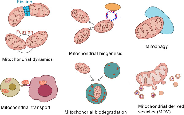
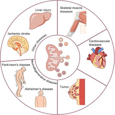
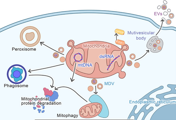
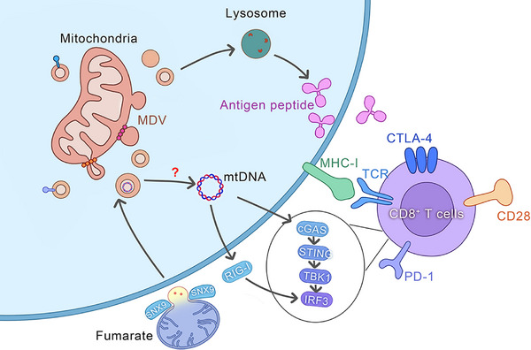
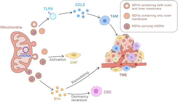
Mitochondria-derived vesicles (MDVs) participate in early cellular defence mechanisms initiated in response to mitochondrial damage. They maintain mitochondrial quality control (MQC) by clearing damaged mitochondrial components, thereby ensuring the normal functioning of cellular processes. This process is crucial for cell survival and health, as mitochondria are the energy factories of cells, and their damage can cause cellular dysfunction and even death. Recent studies have shown that MDVs not only maintain mitochondrial health but also have a significant impact on tumour progression. MDVs selectively encapsulate and transport damaged mitochondrial proteins under oxidative stress and reduce the adverse effects of mitochondrial damage on cells, which may promote the survival and proliferation of tumour cells. Furthermore, it has been indicated that after cells experience mild stress, the number of MDVs significantly increases within 2-6 h, whereas mitophagy, a process of clearing damaged mitochondria, occurs 12-24 h later. This suggests that MDVs play a critical role in the early stress response of cells. Moreover, MDVs also have a significant role in intercellular communication, specifically in the tumour microenvironment. They can carry and transmit various bioactive molecules, such as proteins, nucleic acids, and lipids, which regulate tumour cell's growth, invasion, and metastasis. This intercellular communication may facilitate tumour spread and metastasis, making MDVs a potential therapeutic target. Advances in MDV research have identified novel biomarkers, clarified regulatory mechanisms, and provided evidence for clinical use. These breakthroughs pave the way for novel MDV-targeted therapies, offering improved treatment alternatives for cancer patients. Further research can identify MDVs' role in tumour development and elucidate future cancer treatment horizons.

Keywords: mitochondria‐derived vesicle; target; transport pathway; tumour progression; tumour therapy.


Conflict of interest statement

The authors declare that they have no competing interests.

Figures

FIGURE 1
FIGURE 1
Various mechanisms of mitochondrial quality control (MQC). Mitochondrial quality control comprises a complex network of processes aimed at maintaining mitochondrial health and function. Key components of this control system include mitochondrial dynamics (fission and fusion), mitochondrial biogenesis, mitophagy, mitochondrial trafficking, and mitochondrial proteolysis. The emerging field of mitochondrial‐derived vesicles (MDVs) is pivotal for maintaining mitochondrial homeostasis and cellular health.
FIGURE 2
FIGURE 2
Mitochondrial‐derived vesicles (MDVs) and diseases. MDVs as a mechanism for mitochondrial quality control play a significant role in the development and progression of various diseases, such as tumours, neurodegenerative diseases (such as Parkinson's disease, Alzheimer's disease), cardiovascular diseases, skeletal muscle, etc. Research studies have indicated that MDVs are also involved in the development of diseases such as ischemic stroke and liver injury.
FIGURE 3
FIGURE 3
Transport pathways of mitochondria‐derived vesicles (MDVs). The transport pathways of mitochondrial‐derived vesicles (MDVs) are diverse and complex, mainly consisting of three major routes. First, MDVs can be directionally transported to lysosomes. Second, they can target peroxisomes in a specific manner. Third, MDVs can enter multivesicular bodies (MVBs), then be encapsulated into extracellular vesicles (EVs), and finally be secreted into the extracellular environment. Notably, MDVs can not only precisely target exosomes but also, under specific circumstances, be directed towards phagosomes. Among these pathways, the transport route leading to lysosomes is considered the main one for MDVs. In this process, SNARE‐protein‐mediated vesicle fusion, the endosomal sorting mechanism, and the membrane transport regulation involving the VPS35 protein all play crucial roles. This precise and dynamic transport process not only ensures that MDVs can efficiently and selectively reach specific intracellular destinations but also actively participates in the regulation of various cellular functions and is indispensable for maintaining the stability of the intracellular environment. This multipathway and selective transport mechanism enables MDVs to flexibly respond to various signals inside and outside the cell and participate in a series of important physiological processes, including organelle interaction, substance metabolism, signal transduction, and cell communication. Thus, MDVs play a key role in cellular life activities.
FIGURE 4
FIGURE 4
The mechanisms of mitochondrial‐derived vesicles (MDVs) involved in the mechanism of innate immune response. MDVs play a significant role in modulating innate immune responses. Through their transport of mitochondrial antigens to lysosomes, MDVs present these antigens to CD8+ T cells following processing by lysosomal hydrolases. This process results in the recognition of antigenic peptides on the cell surface, which triggers an immune response by the innate immune system. Another pathway through which MDVs contribute to innate immune responses involves perturbations in mitochondrial morphology and the release of mitochondrial DNA (mtDNA) into the cytoplasm due to fumarate accumulation. This release activates the cGAS–STING–TBK1 pathway, consequently initiating an innate immune response. These intricate mechanisms highlight the pivotal role of MDVs in modulating cellular immune signalling and innate immune defence.
FIGURE 5
FIGURE 5
The mechanisms of mitochondria‐derived vesicles (MDVs) involved in tumour progression. Mitochondrial DNA (mtDNA) serves as a critical cargo within MDVs. Firstly, MDVs carrying mtDNA can activate fibroblasts, leading to the promotion of tumour progression. Secondly, MDVs carrying mtDNA can fuse with exosomes, thereby reshaping the tumour microenvironment. This process can also induce the dormancy reversal of cancer stem cells (CSCs) and facilitate tumour progression. Lastly, MDVs carrying mtDNA can activate Toll‐like receptor 9 (TLR9), resulting in the release of CCL2 which can impact tumour‐associated macrophages (TAMs) and strengthen the immunosuppression within the tumour microenvironment.

Similar articles

References

    1. Jasra IT, Cuesta‐Gomez N, Verhoeff K, Marfil‐Garza BA, Dadheech N, Shapiro AMJ. Mitochondrial regulation in human pluripotent stem cells during reprogramming and beta cell differentiation. Front Endocrinol (Lausanne). 2023;14:1236472. - PMC - PubMed
    1. Rongvaux A. Innate immunity and tolerance toward mitochondria. Mitochondrion. 2018;41:14‐20. - PubMed
    1. Zhang SS, Zhou S, Crowley‐McHattan ZJ, Wang RY, Li JP. A review of the role of endo/sarcoplasmic reticulum‐mitochondria Ca(2+) transport in diseases and skeletal muscle function. Int J Environ Res Public Health. 2021;18. - PMC - PubMed
    1. Amador‐Martínez I, Aparicio‐Trejo OE, Bernabe‐Yepes B, et al. Mitochondrial impairment: a link for inflammatory responses activation in the cardiorenal syndrome Type 4. Int J Mol Sci. 2023;24. - PMC - PubMed
    1. Barabino S, Lombardi S, Zilocchi M. Keep in touch: a perspective on the mitochondrial social network and its implication in health and disease. Cell Death Discov. 2023;9:417. - PMC - PubMed

LinkOut - more resources