. 2025;51(3):e70018.
doi: 10.1111/nan.70018.

Testing Meningiomas With Methylation Arrays: Insights and Recommendations From a Large Single-Centre Study

Affiliations

Testing Meningiomas With Methylation Arrays: Insights and Recommendations From a Large Single-Centre Study

Fernanda Ruiz et al. Neuropathol Appl Neurobiol. 2025.

Abstract

Aims: Meningiomas are common primary CNS tumours, and their morphological diagnosis is usually straightforward. Their histological grading according to CNS WHO criteria alone provides limited information on recurrence risk. Risk stratification of meningiomas combining WHO grade, methylation class and copy number profile improves prediction of the risk of early recurrence. Because of the frequency of meningiomas in diagnostic practice, applying this prediction algorithm to all meningiomas is financially not viable in most healthcare systems.

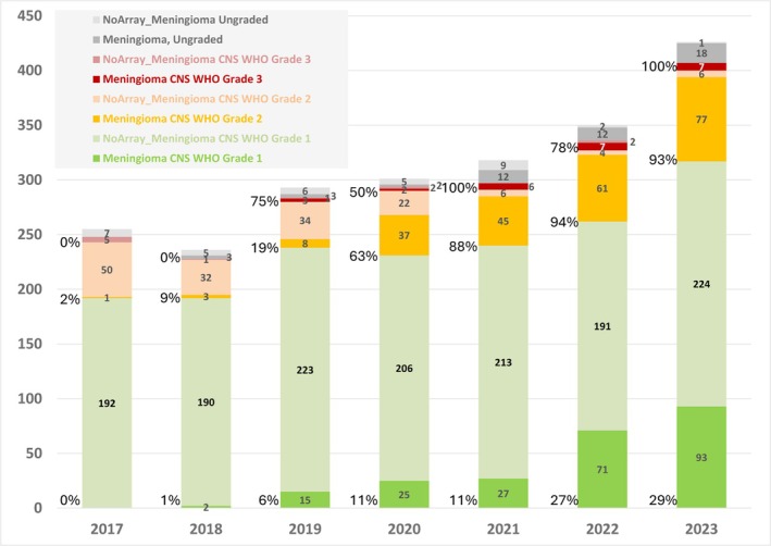
Methods: We analysed a retrospective dataset of over 1000 meningiomas from a single centre with methylation arrays to provide guidance on which meningiomas to prioritise for integrated molecular testing and to understand how WHO grades resolve into risk strata.

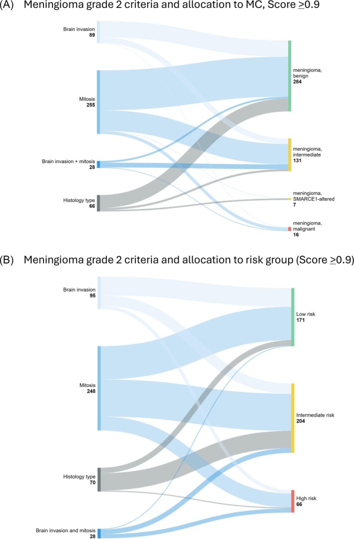
Results: Approximately 90% of CNS WHO Grade 1 meningiomas were allocated into the methylation class 'benign' and also into a low-risk group. Grade 2 meningiomas were allocated almost equally to either the low-risk (39%) or intermediate-risk groups (46%) but occasionally also to the high-risk group (15%). All grading criteria for CNS WHO Grade 2 meningiomas (brain invasion, mitotic count, cytoarchitectural atypia and histological type) showed a similar risk score distribution as the entire group. Grade 3 meningiomas were allocated to intermediate- (26%) or high-risk groups (74%).

Conclusion: Our data suggest that Grade 2 and 3 meningiomas should be prioritised for methylation profiling. A small proportion of Grade 1 meningiomas may also benefit from integrated molecular analysis, and further research is needed to explore if those histologically benign meningiomas with a predicted increased recurrence risk are associated with distinct demographic or histological characteristics.

Keywords: CNS WHO grading; meningioma; methylation array; model score; prognostication.


Conflict of interest statement

Felix Sahm is a co‐founder and shareholder of Heidelberg Epignostix GMBH, which licences the use of the methylation classifier. The other authors declare no conflicts of interest. Felix Sahm, Zane Jaunmuktane and Sebastian Brandner are members of the editorial board at Neuropathology and Applied Neurobiology.

Figures

FIGURE 1
FIGURE 1
Diagnostic workflow in this study. From all locally diagnosed and externally diagnosed meningiomas (n = 2885), a subset (n = 1226) underwent methylation array analysis. Arrays with a calibrated score of 0.9 and higher (n = 990) were used to establish methylation classes. For the calculation of the model score (which requires the WHO grade), the subset of graded meningiomas (n = 924) was included.
FIGURE 2
FIGURE 2
Methylation profiling of meningiomas in the standard diagnostic pathway: 2179 meningiomas were diagnosed between January 2017 and December 2023. The graph indicates the proportion of Grade 1, 2 and 3 meningiomas, as well as ungraded meningiomas. There is a continuous increase of methylation arrays for risk stratification of meningiomas. 29% of Grade 1, 93% of Grade 2 and 100% of Grade 3 meningiomas were profiled in 2023. See also Table S1 for corresponding data.
FIGURE 3
FIGURE 3
(A) Outcome of the methylation profiling of all referred and internally diagnosed meningiomas, with a calibrated score of ≥ 0.9. A majority of Grade 1 tumours are allocated to the methylation class benign, whereas Grade 2 tumours allocate approximately 60% to the MC benign and the remaining tumours into MC of intermediate and rarely malignant. See also Table S3 for corresponding data. (B) Outcome of the methylation profiling of all referred and internally diagnosed meningiomas after calculation of the model score. Of the Grade 1 meningiomas, a significant majority allocate to these three low scores of 0, 1 and 2. Instead, Grade 2 meningiomas show a wider distribution across many model scores courses, underpinning the importance of prioritising these tumours for methylation profiling with risk prediction.
FIGURE 4
FIGURE 4
(A) Stratification of Grade 2 meningiomas by grading criteria (brain invasion, mitotic counts and histology type), and the proportional allocation into MCs benign, intermediate and malignant. Brain invasion and mitotic count show a similar distribution to MCs benign and intermediate, whereas tumours with both increased mitotic count and brain invasion predominantly allocate to the intermediate‐risk group. A majority of meningiomas with histological appearances suggesting Grade 2 were allocated to the benign MC. See also Table 1 for corresponding data. (B) Stratification of Grade 2 meningiomas by grading criteria (brain invasion, mitotic counts and histology type) and the allocation to the risk groups. The criteria brain invasion or mitotic activity distribute similarly into the three risk groups, with histology type predominantly into the intermediate‐risk group. See also Table 2 for corresponding data.
FIGURE 5
FIGURE 5
Stratification of Grade 2 meningiomas by grading criteria (brain invasion, mitotic counts and histology type) and the allocation to the individual model scores. See also Table S6 for corresponding data.
FIGURE 6
FIGURE 6
Contribution of Grade 1, 2 and 3 meningiomas to the group of 1p deleted or 1p intact meningiomas. See also Table S10 for corresponding data.

References

    1. Ostrom Q. T., Price M., Neff C., et al., “CBTRUS Statistical Report: Primary Brain and Other Central Nervous System Tumors Diagnosed in the United States in 2016‐2020,” Neuro‐Oncology 25 (2023): iv1–iv99. - PMC - PubMed
    1. Perry A., Stafford S. L., Scheithauer B. W., Suman V. J., and Lohse C. M., “Meningioma Grading: An Analysis of Histologic Parameters,” American Journal of Surgical Pathology 21 (1997): 1455–1465. - PubMed
    1. Louis D. N., Perry A., Wesseling P., et al., “The 2021 WHO Classification of Tumors of the Central Nervous System: A Summary,” Neuro‐Oncology 23 (2021): 1231–1251. - PMC - PubMed
    1. Wang J. Z., Landry A. P., Raleigh D. R., et al., “Meningioma: International Consortium on Meningiomas (ICOM) Consensus Review on Scientific Advances & Treatment Paradigms for Clinicians, Researchers, and Patients,” Neuro‐Oncology 26, no. 10 (2024): 1742–1780, 10.1093/neuonc/noae082. - DOI - PMC - PubMed
    1. Tauziede‐Espariat A., Parfait B., Besnard A., et al., “Loss of SMARCE1 Expression Is a Specific Diagnostic Marker of Clear Cell Meningioma: A Comprehensive Immunophenotypical and Molecular Analysis,” Brain Pathology 28 (2018): 466–474. - PMC - PubMed

Publication types

LinkOut - more resources