. 2025 Apr 28:13:1453019.
doi: 10.3389/fped.2025.1453019. eCollection 2025.

A report of 12 cases of congenital hepatic hemangioma and literature review

Affiliations

A report of 12 cases of congenital hepatic hemangioma and literature review

Ying Wei et al. Front Pediatr. .

Erratum in

Abstract

Objective: To investigate the clinical features, complications, diagnosis and management of congenital hepatic hemangiomas(CHHs).

Methods: 12 neonates of CHH admitted to our hospital in the past 10 years were retrospectively analyzed, and the clinical manifestations, auxiliary examination results, diagnosis and treatment methods, clinical efficacy andprognosis were reviewed.

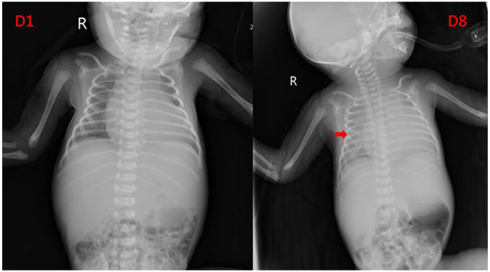
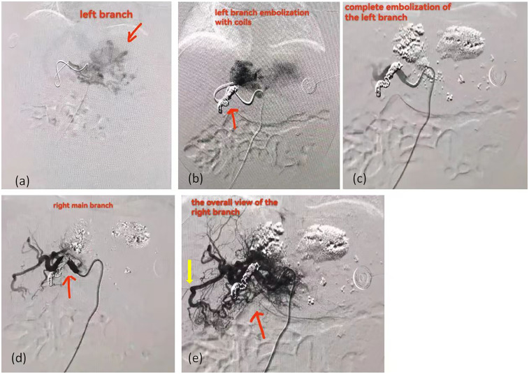
Results: In this study, 12 neonates with CHHs were reported. Among them, 8 cases underwent surgical treatment and recovered well postoperatively. 3 cases received routine pharmacological treatment, were gradually recovering. Only one case, presenting with giant CHH and congestive heart failure (CHF) at birth, failed initial pharmacological treatment and underwent percutaneous hepatic hemangioma embolization but died postoperatively.

Conclusion: Large CHHs tend to be complicated with refractory congestive heart failure, likely due to tumor size and intra-tumor arteriovenous shunt. Propranolol is effective for CHHs with stable hemodynamics but has a slow onset of action, making it less suitable for cases complicated with CHF. Surgical resection is effective and recommended for large CHHs with stable hemodynamics, while percutaneous hepatic hemangioma embolization is advised for unstable cases.

Keywords: congenital hepatic hemangioma; congestive heart failure; percutaneous hepatic hemangioma embolization; propranolol; surgical resection.


Conflict of interest statement

The authors declare that the research was conducted in the absence of any commercial or financial relationships that could be construed as a potential conflict of interest.

Figures

Figure 1
Figure 1
The flowchart of assessment, enrollment and exclusion for the patients.
Figure 2
Figure 2
The ultrasound, CT and pathological images of congenital hepatic hemangioma. (a–d) shows the abdominal enhanced CT images: (a) Arterial phase; (b) Venous phase; (c) Portal phase; (d) Delay phase. The area marked by the arrow is the hemangioma. (e,f) shows the ultrasound images: (e) The two-dimensional gray-scale image; (f) The color Doppler image. The area marked by the arrow is the hemangioma. (g,h) shows the pathological images: (g) The tumor tissue shows varying degrees of vascular proliferation, dilation and congestion, as well as proliferation of fibrous tissue around the vessels. (h) endothelial cells do not express GLUT-1, red blood cells can be used as a positive internal control.
Figure 3
Figure 3
Comparison of chest radiograph before and after pharmacological treatment in case 12. The arrow indicates that the transparency of both lungs is uniformly decreased, suggesting pulmonary edema.
Figure 4
Figure 4
The angiographic images of case 12 received percutaneous hepatic hemangioma embolization. (a) The left branch of the hemangioma. The area marked by the arrow is the hemangioma. (b) The image after embolization of the left main trunk with coils show that there are still hazy malformed vessels at the distal end. The area marked by the arrow is the coils. (c) The image after filling the fine vascular network with ultra-liquefied iodized oil, pingyangmycin, and gelatin sponge particles. (d) The right main trunk of the hemangioma. The area marked by the arrow is the right main trunk. (e) The overall view of right branch of the hemangioma. The red arrow indicates the overall view of right branch; The yellow arrow indicates the arteriovenous shunt.

Similar articles

References

    1. Yan W, Li Z, Yang F, Huang X. Analysis on epidemiological and clinical features of 2761 cases of infantile hemangioma. J Pract Dermatol. (2018) 11(02):71–3. 10.11786/sypfbxzz.1674-1293.20180203 - DOI
    1. Zhao F. Diagnosis and treatment of congenital hemangioma and vascular malformations. Chin J Stomatol. (2005) 40(03):203–5. 10.3760/j.issn:1002-0098.2005.03.008 - DOI - PubMed
    1. Gong X, Yang M, Zhang Z, Qiu T, Zhou J, Shan W, et al. Clinical characteristics and managements of congenital hepatic hemangioma: a cohort study of 211 cases. Hepatol Int. (2024) 40(12):2440. 10.1007/s12072-024-10756-5 - DOI - PubMed
    1. El Zein S, Boccara O, Soupre V, Vieira AF, Bodemer C, Coulomb A, et al. The histopathology of congenital haemangioma and its clinical correlations: a long-term follow-up study of 55 cases. Histopathology. (2020) 77(2):275–83. 10.1111/his.14114 - DOI - PubMed
    1. Iacobas I, Phung TL, Adams DM, Trenor CC, 3rd, Blei F, Fishman DS, et al. Guidance document for hepatic hemangioma (infantile and congenital) evaluation and monitoring. J Pediatr. (2018) 203:294–300.e2. 10.1016/j.jpeds.2018.08.012 - DOI - PubMed

LinkOut - more resources