. 2025 Apr 28:16:1462045.
doi: 10.3389/fimmu.2025.1462045. eCollection 2025.

Independent and temporally separated dynamics for RORγt and Foxp3 during Th17 differentiation

Affiliations

Independent and temporally separated dynamics for RORγt and Foxp3 during Th17 differentiation

Stav Miller et al. Front Immunol. .

Abstract

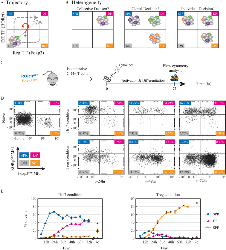
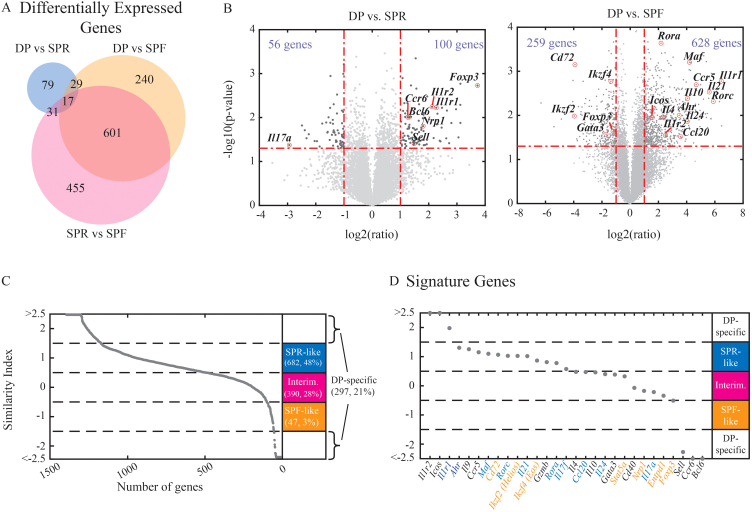
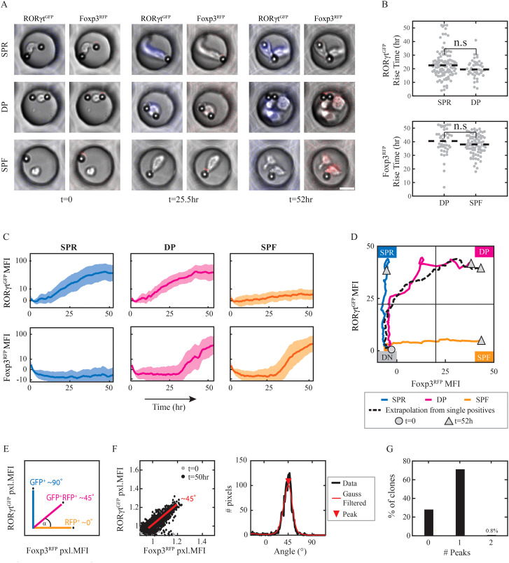
T helper 17 and Regulatory T cells (Th17 and Treg, respectively) are two well-described lymphocyte subsets with opposing actions. The divergent fates of Th17 and Treg cells are accounted for, at least in part, by molecular antagonism that occurs between their respective specific transcription factors, RORγt and Foxp3. An imbalance between Th17 and Treg cells may lead to tissue inflammation and is associated with certain types of autoimmunity. In order to understand the heterogeneity and dynamics of the differentiation process, we studied Th17/Treg cell differentiation of naïve cells in vitro, using RORγtGFPFoxp3RFP dual-reporter mouse. Flow cytometry revealed the consistent emergence of a population of double positive RORγt+Foxp3+ (DP) cells during the early stages of Th17 cell differentiation. These DP cells are closely related to RORγt+ single positive (SPR) cells in terms of global gene expression. Nevertheless, for some genes, DP cells share an expression pattern with Foxp3+ single positive (SPF) Treg cells, most importantly by reducing IL17 levels. Using time-lapse microscopy, we could delineate the expression dynamics of RORγt and Foxp3 at a clonal level. While the RORγt expression level elevates early during differentiation, Foxp3 rises later and is more stable upon environmental changes. These distinct expression profiles are independent of each other. During differentiation and proliferation, individual cells transit between SPR, DP, and SPF states. Nevertheless, the differentiation of sister cells within a clone progeny was highly correlated. We further demonstrated that sorted SPR and DP populations were not significantly affected by changes in their environment, suggesting that the correlated fate decision emerged at early time points before the first division. Overall, this study provides the first quantitative analysis of differentiation dynamics during the generation of DP RORγt+Foxp3+ cells. Characterizing these dynamics and the differentiation trajectory could provide a profound understanding and be used to better define the distinct fates of T cells, critical mediators of the immune response.

Keywords: T-cell differentiation; clonal analysis; double positive T-cells; micro-well array; systems immunology; time-lapse microscopy; transcription factor dynamics.


Conflict of interest statement

Author YEA is a scientific advisory board member and consultant at TeraCyte. The remaining authors declare that the research was conducted in the absence of any commercial or financial relationships that could be construed as a potential conflict of interest.

Figures

Figure 1
Figure 1
A large stable DP population emerges during Th17 cell differentiation. (A) Potential trajectories of naive CD4+ T cells towards expressing both effector and regulatory transcription factors and becoming double positive RORγt+Foxp3+ cells. (B) The fate decision within cells can arise at different levels. The cellular state might be the result of a collective homogeneous decision where all cells become double positive. Alternatively, some cells could attain a double positive fate, while others become single positive. In this case, the decision could occur at the clonal level, such that all the descendants of the same clone have the same fate, or each cell could make an independent individual decision. Cell color represents a clone. (C) Experimental design. Naïve CD4+ T cells were isolated from the spleen of dual reporter RORγtGFPFoxp3RFP mice and cultured in the presence of activation beads and fate-inducing cytokines for 72hr before being analyzed using flow cytometry. (D) Scatter plots showing measured expression patterns of RORγtGFP and Foxp3RFP of naive cells on t=0 and of cells under Th17 (upper panel) and Treg (lower panels) conditions on t=24,48,72hr. Population color code: blue, SPR (single positive, Rorγt); pink, DP (double positive); red, SPF (single positive, Foxp3); gray, DN (double negative). (E) Percentages of detected populations of cells cultured under Th17 (left) and Treg (right) conditions. Measurements were taken every 6hr throughout 72hr and after 7 days using flow cytometry. The presented data is from one representative experiment out of three, each with three technical repeats.
Figure 2
Figure 2
DP cells have a mixed RNA expression profile with reduced immune effector function. Naive cells were activated in the presence of Th17 or Treg-inducing conditions for 3 days and were sorted into SPR (single positive Rorγt+, Th17 condition), DP (double positive Rorγt+Foxp3+, Th17 condition), and SPF (single positive Foxp3+, Treg condition) populations. Bulk RNA was extracted for each sample and used for the construction of the RNA-seq library. (A) Venn diagram showing the number of differentially expressed genes between each pair of populations (fold change above 2 and Pv<0.5, a total of 1416 genes). (B) Volcano plots emphasize changes in specific genes while comparing DP vs. SPR (left) or DP to SPF (right). (C-D) Similarity index between DP to the other populations was calculated. Genes with indices between -1.5 to -0.5 were labeled as “SPF-like” (red), -0.5 to 0.5 as “intermediate” (pink), and 0.5 to 1.5 as “SPR-like” (blue). Genes below -1.5 and above 1.5 are considered to show distinct expression patterns in DP cells. (C) All differentially expressed genes were sorted by their similarity indices. (D) Immune related genes were sorted by their similarity indices (Th17- and Treg-related genes names in blue and red, respectively). An average of three independent biological repeats is presented for each sample.
Figure 3
Figure 3
RORγt and Foxp3 expression dynamics arise at distinct timescales. Naïve CD4+ T cells were plated in micro-wells, supplemented with differentiating cytokines and monitored under the microscope for 52hr. Analysis was done on micro-wells with 1 cell and at least 1 activation bead at the beginning of the experiment. (A) Images of representative micro-wells on t=0,25.5,52hr: elevation of RORγtGFP level in SPR cells under Th17 condition (top), of both RORγtGFP and Foxp3RFP levels in DP cells under Th17 condition (middle) or of Foxp3RFP level in SPF cells under Treg condition (bottom). Scale bar, 10μm. Blue, RORγtGFP+ pixels; Red, Foxp3RFP+ pixels. (B) Fluorescence rise time above response threshold of RORγtGFP in SPR and DP clones under Th17 condition (top); and of Foxp3RFP in DP clones under Th17 condition and in SPF clones under Treg condition (bottom). Dashed line, median values. A two-samples t-test was calculated for all presented pairs, Pv>0.05. Each dot represents one micro-well. (C) Fluorescence intensity over time of RORγtGFP (upper panel) and Foxp3RFP (lower panel) in SPR and DP clones under Th17 condition (left and middle, respectively) and SPF clones under Treg condition (right). A median of data is shown ± std. (D) Median values of traces of SPR, DP, and SPF clones drawn in the space of RORγt (y-axis) vs. Foxp3 (x-axis). Extrapolation of median values of RORγt in SPR clones vs. median values of Foxp3 in SPF clones over time is shown as a dashed line. Circular node, t=0; triangle, t=52hr. (E) Expected pixel angles between RORγtGFP (90°) and Foxp3RFP (0°) depending on fluorescence signals in 3 putative pixels. (F) Pixel fluorescent intensity angles in one representative DP clone: pixel angles on t=0,52hr (left); distribution of pixel angles over last 5 time frames, with Gaussian filtering and maxima local peak (red triangle) at ~45° (right). Each dot represents one pixel. (G) Percentages of micro-wells showing either 0,1 or 2 angles’ peaks at the last 5 time frames. The presented data is from one representative experiment out of two.
Figure 4
Figure 4
Th17 cells’ state is not affected by neighboring cells or environmental cues. (A) Cells were cultured under Th17 and Treg conditions for 2-3 days. Then they were sorted to SPR, DP, and SPF and re-cultured under Th17 or Treg conditions for additional 2 days (day 4 or 5 from the beginning of the experiment). (B) Schematic representation of experimental setup. SPR and DP cells were sorted. Then, SPR cells were stained with eFluor 450 dye to enable their detection post co-culturing. Sorted cells were re-cultured individually (100% SPR; 100% DP) or co-cultured at 3:1 (75% SPR:25% DP) and 1:1 (50% SPR:50% DP) ratios for 2 days. (C, D) Resulting populations (panel of 4 plots) that were SPR originated (eFluor 450+; upper panel) or DP originated (eFluor 450-; lower panel) and re-cultured under Th17 (C) or Treg (D) conditions were measured on flow cytometry. Three independent biological repeats are presented. (E) SPF cells were sorted after 3 days of differentiation and re-cultured under Th17 or Treg condition. Resulting populations were measured on flow cytometry following 48hr of culture. Three independent biological repeats are presented. (F) Scatter plots showing measured expression patterns of RORγtGFP and Foxp3RFP of SPF sorted cells after 2 days of re-culturing under Th17 (left) and Treg (right) conditions. Data is from one representative experiment out of three. Blue, SPR; pink, DP; orange, SPF; white, DN.

Similar articles

References

    1. Korn T, Bettelli E, Oukka M, Kuchroo VK. IL-17 and th17 cells. Annu Rev Immunol. (2009) 27:485–517. doi: 10.1146/annurev.immunol.021908.132710 - DOI - PubMed
    1. Zhu J, Yamane H, Paul WE. Differentiation of effector CD4 T cell populations (*). Annu Rev Immunol. (2010) 28:445–89. doi: 10.1146/annurev-immunol-030409-101212 - DOI - PMC - PubMed
    1. DuPage M, Bluestone JA. Harnessing the plasticity of CD4+ T cells to treat immune-mediated disease. Nat Rev Immunol. (2016) 16:149–63. doi: 10.1038/nri.2015.18 - DOI - PubMed
    1. Antebi YE, Reich-Zeliger S, Hart Y, Mayo A, Eizenberg I, Rimer J, et al. . Mapping differentiation under mixed culture conditions reveals a tunable continuum of T cell fates. PloS Biol. (2013) 11:e1001616. doi: 10.1371/journal.pbio.1001616 - DOI - PMC - PubMed
    1. Eizenberg-Magar I, Rimer J, Zaretsky I, Lara-Astiaso D, Reich-Zeliger S, Friedman N. Diverse continuum of CD4+ T-cell states is determined by hierarchical additive integration of cytokine signals. Proc Natl Acad Sci U S A. (2017) 114:E6447–56. doi: 10.1073/pnas.1615590114 - DOI - PMC - PubMed

MeSH terms

Substances

LinkOut - more resources