A Curious Presentation of a Human Papillomavirus (HPV)-Driven Pelvic Squamous Cell Carcinoma of Unknown Primary: A Case Report
- PMID: 40357111
- PMCID: PMC12068359
- DOI: 10.7759/cureus.82134
A Curious Presentation of a Human Papillomavirus (HPV)-Driven Pelvic Squamous Cell Carcinoma of Unknown Primary: A Case Report
Abstract
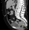
Cancers of unknown primary (CUP) are rare malignancies among invasive cancers and are comprised of a variety of subtypes. In the pelvis, occult squamous cell carcinoma (SCC) usually arises from the vulva, vagina, cervix, or anus. We present the case of a 55-year-old patient with pelvic pain. Magnetic resonance imaging showed a presacral and posterior lower uterine soft tissue mass measuring 6.7 x 5.0 x 6.2 cm with regional lymph node involvement. An exam under anesthesia and colonoscopy did not detect cervical or anal involvement. A biopsy of the mass confirmed poorly differentiated SCC, positive p16 staining on immunohistochemistry, and human papillomavirus (HPV+) RNA in-situ hybridization. The findings were most consistent with an atypical presentation of locally advanced cervical cancer. The patient received chemoradiation with volumetric modulated arc therapy. After four weeks of treatment with some tumor response, she received stereotactic body radiotherapy (SBRT) as the mass was not accessible for brachytherapy. The patient ultimately had a partial response with the shrinking tumor separating from the cervix. She was re-evaluated by a multidisciplinary team, and based on response and imaging, the tumor was most likely determined to originate from the anus. Colorectal surgery assumed care for definitive abdominal perineal resection. This case demonstrates the importance of continuous evaluation of the site of disease in patients with CUP and the avoidance of anchoring on a definitive diagnosis. Identification of the primary site may be impossible initially, but willingness to alter treatment as new information presents is essential to improve patient outcomes.
Keywords: carcinoma of unknown primary (cup); hpv-associated malignancy; human papillomavirus (hpv); occult carcinoma; scc of unknown primary; squamous cell carcinoma (scc); squamous cell rectal carcinoma.
Copyright © 2025, Dau et al.
Conflict of interest statement
Human subjects: Consent for treatment and open access publication was obtained or waived by all participants in this study. Conflicts of interest: In compliance with the ICMJE uniform disclosure form, all authors declare the following: Payment/services info: All authors have declared that no financial support was received from any organization for the submitted work. Financial relationships: All authors have declared that they have no financial relationships at present or within the previous three years with any organizations that might have an interest in the submitted work. Other relationships: All authors have declared that there are no other relationships or activities that could appear to have influenced the submitted work.
Figures


References
-
- NCCN clinical practice guidelines occult primary. Ettinger DS, Agulnik M, Cates JM, et al. J Natl Compr Canc Netw. 2011;9:1358–1395. - PubMed
-
- Cancer of unknown primary: biological and clinical characteristics. Pavlidis N. Ann Oncol. 2003;14 Suppl 3:0–8. - PubMed
-
- Cancers linked with HPV each year. [ May; 2024 ]. 2024. https://www.cdc.gov/cancer/hpv/cases.html https://www.cdc.gov/cancer/hpv/cases.html
-
- NCCN Clinical Practice Guidelines in Oncology (NCCN Guidelines®) Cervical Cancer. Accessed. [ Jul; 2024 ]. https://www.nccn.org/professionals/physician_gls/pdf/cervical.pdf https://www.nccn.org/professionals/physician_gls/pdf/cervical.pdf
Publication types
LinkOut - more resources
Full Text Sources
Research Materials
