. 2025 Apr 28:13:1575341.
doi: 10.3389/fbioe.2025.1575341. eCollection 2025.

Structural and functional analysis of a homotrimeric collagen peptide

Affiliations

Structural and functional analysis of a homotrimeric collagen peptide

Xinling Zhang et al. Front Bioeng Biotechnol. .

Abstract

Objective: This study aimed to chemically synthesize a homotrimeric collagen peptide, evaluate its safety, and assess its effectiveness in promoting collagen synthesis.

Methods: A homotrimeric collagen peptide was synthesized and structurally characterized using circular dichroism and infrared spectroscopy. Thermal stability was analyzed by TG-DSC, and molecular weight and amino acid composition were determined. In vitro cytotoxicity testing assessed safety, while UV-induced photoaging experiments evaluated its effects on collagen and elastin synthesis. In vivo studies in BALB/c mice examined its impact on collagen content, skin structure, and angiogenesis.

Results: The synthesized collagen peptide exhibited high purity (99.1%) and an amino acid composition of glycine, proline, and hydroxyproline in a balanced ratio (15:17:13). Structural analysis confirmed a stable triple-helical conformation similar to type I collagen with excellent thermal stability (Tm = 326.15°C). Cytotoxicity testing showed no adverse effects on cell viability. In vitro, the peptide significantly enhanced collagen and elastin synthesis in fibroblasts. In vivo, intradermal and subcutaneous injection increased collagen content, improved skin structure, and enhanced microvessel density.

Conclusion: This study presents a chemically synthesized homotrimeric collagen peptide with superior purity, structural stability, and biological efficacy in promoting collagen synthesis. Compared to previous studies, this biomimetic material exhibits exceptional thermal stability (Tm = 326.15°C) and a well-balanced amino acid composition, enabling applications in cosmetics and medical devices requiring heat sterilization (e.g., autoclaving), as validated by our patented method (China Patent No. ZL202410309842.9).

Keywords: collagen peptides; heat stability; homotrimer; safety; synthesis.


Conflict of interest statement

Authors KL, TT, BZ, YL, and XQ were employed by LivingPhoenix Regenerative Technologies Development (Chengdu) Co., Ltd. The remaining authors declare that the research was conducted in the absence of any commercial or financial relationships that could be construed as a potential conflict of interest.

Figures
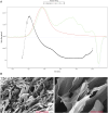
FIGURE 1
FIGURE 1
Gel Permeation Chromatography (GPC) analysis of collagen peptide molecular weight and scanning electron microscopy images. (A) Molecular weight of collagen peptide. Note: Mw stands for weight-average molecular weight, Mn for number-average molecular weight, Mp for peak molecular weight, and PD for polydispersity index. A higher value indicates a broader molecular weight distribution. (B) Scanning electron microscopy images of collagen peptide. Left: Displays the surface morphology of the collagen peptide, revealing its fibrous structure. The surface appears rough, with numerous pores and irregular features. Right: Provides a higher-magnification view of the collagen peptide, offering a more detailed look at its surface characteristics. The image highlights the intricate intertwining and interweaving of collagen fibers, forming a complex three-dimensional network.
FIGURE 2
FIGURE 2
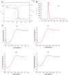
Infrared spectroscopy analysis of the molecular structural characteristics of collagen peptides. Reference to bovine type I collagen protein. Top Image: Infrared (IR) spectrum of the collagen peptide, showing its absorption peaks at different wavenumbers. Middle Image: Infrared (IR) spectrum of bovine type I collagen, displaying its absorption peaks at various wavenumbers. Bottom Image: Comparative infrared (IR) spectra of collagen peptide and bovine type I collagen. Blue curve: IR spectrum of the collagen peptide. Red curve, Red curve: IR spectrum of bovine type I collagen. The two samples exhibit similarities in the positions and intensities of their major absorption peaks, indicating that the collagen peptide shares molecular structural characteristics with bovine type I collagen.
FIGURE 3
FIGURE 3
Detection of thermal stability of collagen peptides. (A) Analysis of the thermal stability of collagen peptides using TG and DSC methods. (B) Analysis of bovine type I collagen protein (as reference) using DSC method. (C) Assessment of the thermal stability of collagen peptides using circular dichroism spectroscopy at 20°C, 40°C, 60°C, and 80°C. Note: TG: Thermogravimetric Analysis; DSC: Differential Scanning Calorimetry.
FIGURE 4
FIGURE 4
Collagen peptides promote collagen synthesis in vitro (A). Cytotoxicity of collagen peptides at different concentrations. (B) The relative mRNA expression levels of type I collagen (col1a1) gene and type III collagen (col3a1) gene. (C) Collagen peptides can enhance the gene expression of col1a1, col3a1, and elastin in cells after ultraviolet radiation damage. Note: * indicates p-value <0.05; ** indicates p-value <0.01; *** indicates p-value <0.001.
FIGURE 5
FIGURE 5
Collagen peptides can improve the structure of mouse skin and promote proliferation of the dermal layer. (A) HE-stained images of mouse skin tissue one and 4 weeks after collagen peptide injection. (B) Masson-stained images of mouse skin tissue one and 4 weeks after collagen peptide injection.
FIGURE 6
FIGURE 6
Injection of collagen peptides in vivo promotes collagen synthesis in mouse skin. Up image, COL1 expression in mouse skin tissue; Down image, COL3 expression in mouse skin tissue. Magnified views of specific regions showing detailed. Blue squares indicate areas of interest with higher magnification.
FIGURE 7
FIGURE 7
Observation of collagen in the dermal layer of mouse skin after collagen protein injection using second harmonic generation microscopy. Sparse collagen fibers with low density and limited network formation at all time points, with minimal structural changes over 4 weeks in control group. Increased collagen density and network formation at 1 week post-injection, with progressive enhancement in collagen fiber density and organization over time. By 4 weeks, a well-defined, dense collagen network was observed in study group.
FIGURE 8
FIGURE 8
In vivo injection of collagen peptides promotes neovascularization in mouse skin. Immunohistochemical images of CD31 in skin tissue after one and 4 weeks of collagen peptide injection (up). Signal quantification graph in the immunohistochemical images of CD31 (down). Note: ns indicates p-value >0.05; * indicates p-value <0.05.

References

    1. Acke F. R., Dhooge I. J., Malfait F., De Leenheer E. M. (2012). Hearing impairment in Stickler syndrome: a systematic review. Orphanet J. Rare Dis. 7, 84. 10.1186/1750-1172-7-84 - DOI - PMC - PubMed
    1. An B., Lin Y. S., Brodsky B. (2016). Collagen interactions: drug design and delivery. Adv. Drug Deliv. Rev. 97, 69–84. 10.1016/j.addr.2015.11.013 - DOI - PubMed
    1. Avila Rodríguez M. I., Rodríguez Barroso L. G., Sánchez M. L. (2018). Collagen: a review on its sources and potential cosmetic applications. J. Cosmet. Dermatol 17 (1), 20–26. 10.1111/jocd.12450 - DOI - PubMed
    1. Badylak S. F., Freytes D. O., Gilbert T. W. (2015). Reprint of: extracellular matrix as a biological scaffold material: structure and function. Acta Biomater. 23 (Suppl. l), S17–S26. 10.1016/j.actbio.2015.07.016 - DOI - PubMed
    1. Bhatwa A., Wang W., Hassan Y. I., Abraham N., Li X.-Z., Zhou T. (2021). Challenges associated with the formation of recombinant protein inclusion bodies in Escherichia coli and strategies to address them for industrial applications. Front. Bioeng. Biotechnol. 9, 630551. 10.3389/fbioe.2021.630551 - DOI - PMC - PubMed

LinkOut - more resources