. 2025 Apr 29;14(9):651.
doi: 10.3390/cells14090651.

Multipotent Mesenchymal Stem Cell Therapy for Vascular Dementia

Affiliations

Multipotent Mesenchymal Stem Cell Therapy for Vascular Dementia

Eun-Young Kim et al. Cells. .

Abstract

Vascular dementia (VD), characterized by cognitive decline and behavioral disorders, has seen a rapid increase in prevalence in recent years. However, effective treatments for VD remain unavailable. Due to its regenerative potential, stem cell therapy has garnered attention as a promising approach for VD treatment, yet it has shown limited effects on cognitive and behavioral impairments caused by the disease. To address this limitation, this study aimed to develop a novel treatment using human embryonic stem cell-derived multipotent mesenchymal stem cells (MMSCs). The therapeutic efficacy of MMSCs was evaluated using a vascular dementia mouse model induced by bilateral carotid artery stenosis (BCAS). The effects of MMSCs were assessed through behavioral tests and postmortem brain tissue analysis, including mRNA expression analysis and hematoxylin and eosin (H&E) staining. MMSCs treatment significantly improved both working memory and long-term memory. Histological analysis revealed enhanced angiogenesis, preservation of blood-brain barrier integrity, and improved hippocampal organization. Furthermore, MMSCs treatment reduced the expression of Rock1/2, indicating suppression of neuroinflammatory and apoptotic pathways. These findings suggest that MMSCs offer a sustainable and effective therapeutic approach for vascular dementia.

Keywords: carotid stenosis; cognitive dysfunction; conduct disorder; human embryonic stem cells; stem cells; vascular dementia.


Conflict of interest statement

Authors Eun-Young Kim, Ki-Sung Hong, Hyung-Min Chung, Se-Pill Park were employed by the company Miraecellbio Co., Ltd. The remaining authors declare that the research was conducted in the absence of any commercial or financial relationships that could be construed as a potential conflict of interest. The company had no role in the design of the study; in the collection, analyses, or interpretation of data; in the writing of the manuscript, or in the decision to publish the results.

Figures
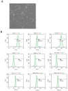
Figure 1
Figure 1
Morphological characteristics, immunophenotyping, and in vivo tracking of MMSCs. (A) Morphology of MMSCs: Light microscopy image at 200× magnification showing the typical fibroblast-like spindle shape of MMSCs. (B) Immunophenotyping results of MMSCs: MMSCs were stained with monoclonal antibodies conjugated to PE and FITC against CD73, CD90, CD105, CD34, CD45, HLA-DR, OCT3/4, TRA-1-60, and TRA-1-81. Green histograms represent the reactivity of the antibodies, and the bars indicate the ratio of antibody reactivity to the background signal. FACS analysis demonstrated positive expressions of CD73, CD90, and CD105, while CD34, CD45, HLA-DR, OCT3/4, TRA-1-60, and TRA-1-81 were negative.
Figure 2
Figure 2
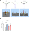
Summary of the Y-maze test. (A) Movement paths of each group overlapped in the Y-maze experiment for working memory measurement. (B) Alternation (%) in the Y-maze test. Data are presented as mean ± SEM. **** p < 0.001. NT: normal type; VD: vascular dementia; MMSCs: multipotent mesenchymal stem cells; BMSCs: bone marrow-derived mesenchymal stem cells; Done: donepezil.
Figure 3
Figure 3
Results of the Barnes maze test and passive avoidance test. (A) Visualization of movement paths for each group during the Barnes maze test. (B) Latency to target for each group across days in the Barnes maze test. (C) Time spent in the target quadrant (TSTQ) as a percentage of total activity time, calculated as the time spent in the target quadrant divided by the total activity time, for each group in the Barnes maze test. Data are presented as mean ± SEM. (D) Entry latency (sec) into the dark compartment during the passive avoidance test across days. **** p < 0.0001 NT: normal type; VD: vascular dementia; MMSCs: multipotent mesenchymal stem cells; BMSCs: bone marrow-derived mesenchymal stem cells; Done: donepezil.
Figure 4
Figure 4
Results of the open field test and rotarod test. (A) Visualization of movement paths of each group in the open field chamber. (B) Graphs showing the total distance traveled (cm) and time spent in the peripheral zone (%) by each group during the open field test. Data are presented as mean ± SEM. (C) Graphs representing the latency to fall down (sec) and RPM recorded for each group during the rotarod test. **** p < 0.0001. NT: normal type; VD: vascular dementia; MMSCs: multipotent mesenchymal stem cells; BMSCs: bone marrow-derived mesenchymal stem cells; Done: donepezil.
Figure 5
Figure 5
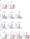
mRNA expression levels analyzed from brain tissues of mice in each group. (A) Genes associated with the ROCK pathway, (B) genes related to the BBB, (C) genes involved in angiogenesis, and (D) genes associated with neuronal and glial cells. All graphs are presented as mean ± standard deviation (SD). * p < 0.05; ** p < 0.01; *** p < 0.001; **** p < 0.0001. NT: normal type; VD: vascular dementia; MMSCs: multipotent mesenchymal stem cells; BMSCs: bone marrow-derived mesenchymal stem cells; Done: donepezil.
Figure 6
Figure 6
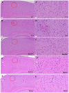
H&E staining of brain sections from each group of mice, observed at 200× magnification. The images show the CA3 and dentate gyrus (DG) regions of the hippocampus. Nuclei were stained purple, and cytoplasm was stained pink. (A) NT group, (B) VD group, (C) MMSC group, (D) BMSC group, and (E) Done group. NT: normal type; VD: vascular dementia; MMSC: multipotent mesenchymal stem cell; BMSC: bone marrow-derived mesenchymal stem cell; Done: donepezil. Representative images from n = 5 mice, with 4–5 sections per mouse analyzed.
Figure 7
Figure 7
The morphology and structure of neurons in cortex of mouses, HE×100 (Left A–E) or 400 (Right A–E). (A) NT group, (B) BCAS group, (C) MMSC (D) BMSC group, and (E) Done group. Irregular neuron arrangement, pyknotic and dark nuclei, and a large number of vacuoles appeared in the VD group, and normal neuron arrangement and nuclei were observed in the MMSC group. (Red circle) However, nuclear condensation similar to VD group was observed in the BMSC group, and a large number of vacuole were observed in the Done group. Representative images from n = 5 mice, with 4–5 sections per mouse analyzed.

References

    1. Iemolo F., Duro G., Rizzo C., Castiglia L., Hachinski V., Caruso C. Pathophysiology of vascular dementia. Immun. Ageing. 2009;6:13. doi: 10.1186/1742-4933-6-13. - DOI - PMC - PubMed
    1. Griffiths A., Matu J., Tang E.Y.H., Gregory S., Anderson E., Fairley A., Townsend R., Stevenson E., Stephan B.C.M., Siervo M., et al. Foods, dietary patterns, and risk of vascular dementia: A systematic review. Nutr. Metab. 2024;21:105. doi: 10.1186/s12986-024-00880-2. - DOI - PMC - PubMed
    1. Jellinger K.A. Pathology and pathogenesis of vascular cognitive impairment-a critical update. Front. Aging Neurosci. 2013;5:17. doi: 10.3389/fnagi.2013.00017. - DOI - PMC - PubMed
    1. Bir S.C., Khan M.W., Javalkar V., Toledo E.G., Kelley R.E. Emerging Concepts in Vascular Dementia: A Review. J. Stroke Cerebrovasc. Dis. 2021;30:105864. doi: 10.1016/j.jstrokecerebrovasdis.2021.105864. - DOI - PubMed
    1. Battle C.E., Abdul-Rahim A.H., Shenkin S.D., Hewitt J., Quinn T.J. Cholinesterase inhibitors for vascular dementia and other vascular cognitive impairments: A network meta-analysis. Cochrane Database Syst. Rev. 2021;2:CD013306. doi: 10.1002/14651858.CD013306. - DOI - PMC - PubMed

Publication types

LinkOut - more resources