

Iron Metabolism and Muscle Aging: Where Ferritinophagy Meets Mitochondrial Quality Control
- PMID: 40358196
- PMCID: PMC12072144
- DOI: 10.3390/cells14090672
Iron Metabolism and Muscle Aging: Where Ferritinophagy Meets Mitochondrial Quality Control
Abstract
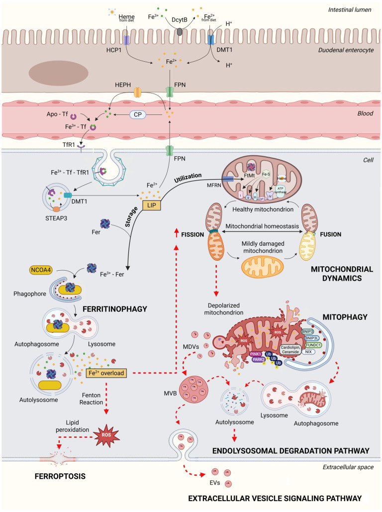
In older adults with reduced physical performance, an increase in the labile iron pool within skeletal muscle is observed. This accumulation is associated with an altered expression of mitochondrial quality control (MQC) markers and increased mitochondrial DNA damage, supporting the hypothesis that impaired MQC contributes to muscle dysfunction during aging. The autophagy-lysosome system plays a critical role in MQC by tagging and engulfing proteins and organelles for degradation in lysosomes. The endolysosomal system is also instrumental in transferrin recycling, which, in turn, regulates cellular iron uptake. In the neuromuscular system, the autophagy-lysosome system supports the structural integrity of neuromuscular junctions, and its dysfunction contributes to muscle atrophy. While MQC was thought to protect against iron-induced cell death, the discovery of ferroptosis, a form of iron-dependent cell death, has highlighted a complex interplay between MQC and iron-inflicted damage. Ferritinophagy, the autophagic degradation of ferritin, if overactivated, can induce ferroptosis. Alternatively, aging may impair ferritinophagy, leading to ferritin accumulation and the release of toxic labile iron under stress, exacerbating oxidative damage and cellular senescence. Physical activity supports muscle health also by preserving mitochondrial quantity and quality and enhancing bioenergetics. However, therapeutic strategies for preventing or reversing physical function decline in aging are still lacking due to the insufficient understanding of the underlying mechanisms. Unveiling how disruptions in iron homeostasis impact muscle quality in older adults may allow for the development of therapeutic strategies targeting iron handling to alleviate age-associated muscle decline.
Keywords: autophagy; cytokine; endolysosomal system; hepcidin; inflammation; labile iron; mitophagy; physical performance; sarcopenia; transferrin.
Conflict of interest statement
The authors declare no conflicts of interest.
Figures
References
-
- Picca A., Triolo M., Wohlgemuth S.E., Martenson M.S., Mankowski R.T., Anton S.D., Marzetti E., Leeuwenburgh C., Hood D.A. Relationship between mitochondrial quality control markers, lower extremity tissue composition, and physical performance in physically inactive older adults. Cells. 2023;12:183. doi: 10.3390/cells12010183. - DOI - PMC - PubMed
-
- Picca A., Mankowski R.T., Kamenov G., Anton S.D., Manini T.M., Buford T.W., Saini S.K., Calvani R., Landi F., Bernabei R., et al. Advanced age is associated with iron dyshomeostasis and mitochondrial DNA damage in human skeletal muscle. Cells. 2019;8:1525. doi: 10.3390/cells8121525. - DOI - PMC - PubMed
-
- Picca A., Saini S.K., Mankowski R.T., Kamenov G., Anton S.D., Manini T.M., Buford T.W., Wohlgemuth S.E., Xiao R., Calvani R., et al. Altered expression of mitoferrin and frataxin, larger labile iron pool and greater mitochondrial DNA damage in the skeletal muscle of older adults. Cells. 2020;9:2579. doi: 10.3390/cells9122579. - DOI - PMC - PubMed
Publication types
MeSH terms
Substances
Grants and funding
LinkOut - more resources
Full Text Sources
Medical
