. 2025 Jun 17;99(6):e0065625.
doi: 10.1128/jvi.00656-25. Epub 2025 May 13.

The NSP6-L260F substitution in SARS-CoV-2 BQ.1.1 and XBB.1.16 lineages compensates for the reduced viral polymerase activity caused by mutations in NSP13 and NSP14

Affiliations

The NSP6-L260F substitution in SARS-CoV-2 BQ.1.1 and XBB.1.16 lineages compensates for the reduced viral polymerase activity caused by mutations in NSP13 and NSP14

Yuri Furusawa et al. J Virol. .

Abstract

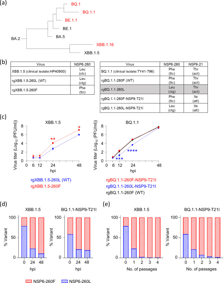
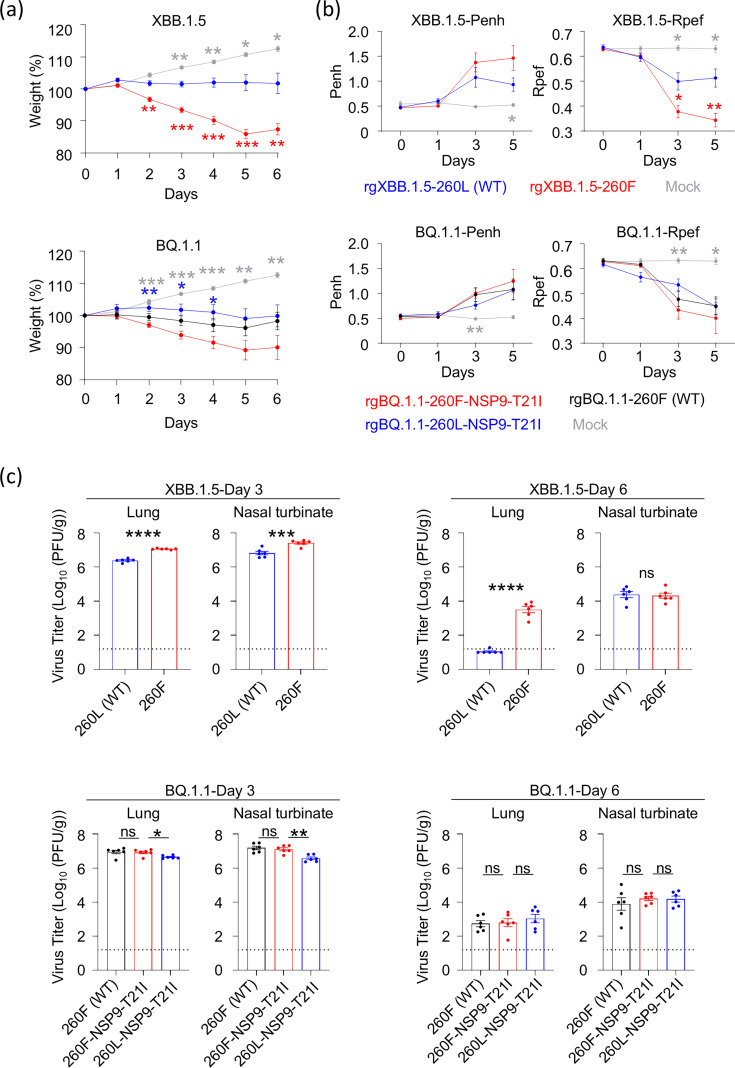
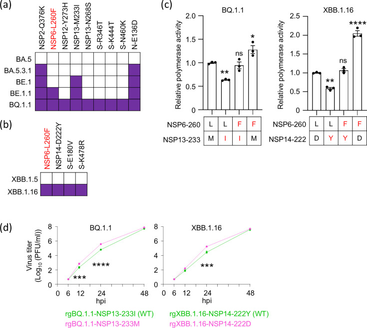
Severe acute respiratory syndrome coronavirus 2 (SARS-CoV-2) Omicron variants emerged at the end of 2021, and their subvariants are still circulating worldwide. While changes in the S protein of these variants have been extensively studied, the roles of amino acid substitutions in non-structural proteins have not been fully revealed. In this study, we found that SARS-CoV-2 bearing the NSP6-L260F substitution emerged repeatedly when we generated several SARS-CoV-2 variants by reverse genetics or when we passaged SARS-CoV-2 isolated from clinical samples and that it was selected under cell culture conditions. Although this substitution has been detected in BQ.1.1 and XBB.1.16 that circulated in nature, its effect on viral properties is unclear. Here, we generated SARS-CoV-2 with or without the NSP6-L260F by reverse genetics and found that NSP6-L260F promotes virus replication in vitro and in vivo by increasing viral polymerase activity and enhancing virus pathogenicity in hamsters. We also identified disadvantageous substitutions, NSP13-M233I and NSP14-D222Y, that reduced BQ.1.1 and XBB.1.16 replication, respectively. These adverse effects were compensated for by NSP6-L260F. Our findings suggest the importance of NSP6-L260F for virus replication and pathogenicity and reveal part of the evolutionary process of Omicron variants.IMPORTANCEAlthough the properties of severe acute respiratory syndrome coronavirus 2 (SARS-CoV-2) Omicron variants continue to change through the acquisition of various amino acid substitutions, the roles of the amino acid substitutions in the non-structural proteins have not been fully explored. In this study, we found that the NSP6-L260F substitution enhances viral polymerase activity and is important for viral replication and pathogenicity. In addition, we found that the NSP13-M233I substitution in the BQ.1.1 lineage and the NSP14-D222Y substitution in the XBB.1.16 lineage reduce viral polymerase activity, and this adverse effect is compensated for by the NSP6-L260F substitution. Our results provide insight into the evolutionary process of SARS-CoV-2.

Keywords: COVID-19; SARS-CoV-2.


Conflict of interest statement

The authors declare no conflict of interest.

Figures

Fig 1
Fig 1
The effect of NSP6-L260F on the growth kinetics of SARS-CoV-2. (a) Phylogenetic tree of SARS-CoV-2 viruses. Lineages with the NSP6-L260F substitution are shown in red. (b) The amino acid and nucleotide sequences of the clinical isolate and viruses generated by reverse genetics are shown. We used a different codon for NSP6-260L compared to the clinical isolate to reduce the potential for reversion. As clonal rgBQ.1.1-260L (highlighted in gray) could not be rescued, we used BQ.1.1 viruses possessing NSP9-T21I in addition to the NSP6-L260F substitution for subsequent experiments. (c) VeroE6/TMPRSS2 cells were infected with each virus at a multiplicity of infection (MOI) of 0.0001. Virus titers at the indicated time points were determined by using plaque assays (n = 3, mean ± s.e.m.). (d and e) rgXBB.1.5-260L (WT) and rgXBB.1.5-260F or rgBQ.1.1-260F-NSP9-T21I and rgBQ.1.1-260L-NSP9-T21I were mixed at an equal ratio on the basis of their infectious titers, and the virus mixture was infected into VeroE6/TMPRSS2 cells grown on 24-well plates in triplicate at an MOI of 0.001. At the indicated time points or at each passage, the proportion of each virus was determined by deep sequencing analysis. Data were analyzed by using a two-way ANOVA with Dunnett’s multiple comparisons test. Statistical significance was calculated against the values in rgXBB.1.5-WT- or rgBQ.1.1-260F-NSP9-T21I-infected cells. ∗P < 0.05, ∗∗P < 0.01, ∗∗∗P < 0.001, and ∗∗∗∗P < 0.0001.
Fig 2
Fig 2
The effect of NSP6-L260F on viral in vivo characteristics. Syrian hamsters were intranasally inoculated with 105 PFU (in 100 µL) of the indicated virus. (a) Body weights of virus-infected and mock-infected hamsters (n = 5) were monitored daily for 6 days. Data are presented as the mean percentages of the starting weight (± s.e.m.). (b) Pulmonary function analyses in infected hamsters. Penh and Rpef were measured by using whole-body plethysmography (n = 5, mean ± s.e.m.). (c) Virus titers in infected Syrian hamsters. Hamsters (n = 5) were euthanized at 3 and 6 days post-infection for virus titration. Virus titers in the nasal turbinate and lungs were determined by using plaque assays. Vertical bars show the mean ± s.e.m. Points indicate data from individual hamsters. The lower limit of detection is indicated by the horizontal dashed line. Data were analyzed by using a two-way ANOVA with Dunnett’s multiple comparisons test (a and b), t-test (c, XBB.1.5-backbone), or a one-way ANOVA with Dunnett’s multiple comparisons test (c, BQ.1.1-backbone). Statistical significance between rgXBB.1.5-260L (WT) and rgXBB.1.5-260F is shown by red asterisks, that between rgBQ.1.1-260F-NSP9-T21I and rgBQ.1.1-260L-NSP9-T21I by blue asterisks, and that between rgXBB.1.5-260L (WT) or rgBQ.1.1-260F-NSP9-T21I and mock by gray asterisks. ∗P < 0.05, ∗∗P < 0.01, ∗∗∗P < 0.001, ∗∗∗∗P < 0.0001, and ns; not significant.
Fig 3
Fig 3
The role of NSP6-L260F in virus transcription/replication. (a) VeroE6/TMPRSS2 cells were infected with each virus at an MOI of 1. At the indicated time points, total RNA was extracted from the infected cells, and the relative expression of sgRNA for the E gene was measured by RT-qPCR (n = 3, mean ± s.e.m.). Data were analyzed by using a two-way ANOVA with Dunnett’s multiple comparisons test. Statistical significance between rgXBB.1.5-260L (WT) and rgXBB.1.5-260F is shown by red asterisks, that between rgBQ.1.1-260F-NSP9-T21I and rgBQ.1.1-260L-NSP9-T21I by blue asterisks, and that between rgBQ.1.1-260F-NSP9-T21I and rgBQ.1.1-260F (WT) by black asterisks. (b) Polymerase activity in HEK293T cells transfected with the replicative cDNA containing the nanoluciferase gene was determined at 72 h post-transfection (n = 3, mean ± s.e.m.). The results are representative of three independent experiments. Data were analyzed by using a t-test. ∗P < 0.05, ∗∗P < 0.01, ∗∗∗P < 0.001, and ∗∗∗∗P < 0.0001.
Fig 4
Fig 4
The role of NSP6-L260F in IFN responses. (a) NSP6 inhibition of RIG-I-dependent IFN-β production. HEK293T cells were transfected with the indicated viral protein expression plasmids, p125-luc, and pGL4.74 [hRluc/TK] vector, with or without a plasmid encoding the constitutively active mutant N-Myc-RIG-IN. IFN-β promoter activity was calculated by normalizing the firefly luciferase activity to the Renilla luciferase activity. The IFN-β promoter activity without N-Myc-RIG-IN was set to 1. The data are shown as mean relative IFN-β promoter activities (n = 3, mean ± s.e.m.). (b) NSP6 inhibition of type-I IFN signaling. HEK293T cells were transfected with the indicated viral protein expression plasmids, pISRE-luc, and pGL4.74 [hRluc/TK] vector. Eight hours after treatment with IFN-α, firefly and Renilla luciferase activities were measured by using a dual-luciferase assay. ISRE-driven firefly luciferase activity was calculated by normalization to the Renilla luciferase activity. The firefly luciferase activity without IFN-α was set to 1. The data are shown as mean relative firefly luciferase activities (n = 3, mean ± s.e.m.). NSP6 expression was confirmed by western blotting. (a and b) The results are representative of three independent experiments. Data were analyzed by using a one-way ANOVA with Dunnett’s multiple comparisons test. ∗∗∗∗P < 0.0001, ns; not significant.
Fig 5
Fig 5
Identification of disadvantageous mutations that are compensated for by NSP6-L260F. (a and b) Amino acid residues that differ between the indicated lineages. (c) The effect of amino acid combinations on polymerase activity. Polymerase activity in HEK293T cells transfected with the replicon DNA containing the nanoluciferase gene was determined at 72 h post-transfection (n = 3, mean ± s.e.m.). Amino acids that are the same as the prototype are shown in black, and mutations that BQ.1.1 or XBB.1.16 carry are shown in red. Data were analyzed by using a one-way ANOVA with Dunnett’s multiple comparisons test. Statistical significance was calculated against the values when the amino acids in both positions were the same as in the prototype. ∗P < 0.05, ∗∗P < 0.01, ∗∗∗P < 0.001, ∗∗∗∗P < 0.0001, and ns, not significant. The results are representative of three independent experiments. (d) VeroE6/TMPRSS2 cells were infected with the indicated virus at an MOI of 0.0001. Virus titers at the indicated time points were determined by using plaque assays (n = 3, mean ± s.e.m.). Data were analyzed by using a two-way ANOVA with Dunnett’s multiple comparisons test. ∗∗∗P < 0.001 and ∗∗∗∗P < 0.0001.

Similar articles

References

    1. Zhu N, Zhang D, Wang W, Li X, Yang B, Song J, Zhao X, Huang B, Shi W, Lu R, Niu P, Zhan F, Ma X, Wang D, Xu W, Wu G, Gao GF, Tan W, China Novel Coronavirus Investigating and Research Team . 2020. A novel coronavirus from patients with pneumonia in China, 2019. N Engl J Med 382:727–733. doi: 10.1056/NEJMoa2001017 - DOI - PMC - PubMed
    1. Huang C, Wang Y, Li X, Ren L, Zhao J, Hu Y, Zhang L, Fan G, Xu J, Gu X, et al. 2020. Clinical features of patients infected with 2019 novel coronavirus in Wuhan, China. The Lancet 395:497–506. doi: 10.1016/S0140-6736(20)30183-5 - DOI - PMC - PubMed
    1. Malone B, Urakova N, Snijder EJ, Campbell EA. 2022. Structures and functions of coronavirus replication-transcription complexes and their relevance for SARS-CoV-2 drug design. Nat Rev Mol Cell Biol 23:21–39. doi: 10.1038/s41580-021-00432-z - DOI - PMC - PubMed
    1. Ricciardi S, Guarino AM, Giaquinto L, Polishchuk EV, Santoro M, Di Tullio G, Wilson C, Panariello F, Soares VC, Dias SSG, Santos JC, Souza TML, Fusco G, Viscardi M, Brandi S, Bozza PT, Polishchuk RS, Venditti R, De Matteis MA. 2022. The role of NSP6 in the biogenesis of the SARS-CoV-2 replication organelle. Nature 606:761–768. doi: 10.1038/s41586-022-04835-6 - DOI - PMC - PubMed
    1. Bills C, Xie X, Shi P-Y. 2023. The multiple roles of nsp6 in the molecular pathogenesis of SARS-CoV-2. Antiviral Res 213:105590. doi: 10.1016/j.antiviral.2023.105590 - DOI - PMC - PubMed

Substances

Supplementary concepts

LinkOut - more resources