. 2025 Aug;14(8):393-408.
doi: 10.1089/wound.2024.0291. Epub 2025 May 13.

SEMTWIST Quantification of Biofilm Infection in Human Chronic Wound Using Scanning Electron Microscopy and Machine Learning

Affiliations

SEMTWIST Quantification of Biofilm Infection in Human Chronic Wound Using Scanning Electron Microscopy and Machine Learning

Surabhi Singh et al. Adv Wound Care (New Rochelle). 2025 Aug.

Abstract

Objective: To develop scanning electron microscopy-based Trainable Weka (Waikato Environment for Knowledge Analysis) Intelligent Segmentation Technology (SEMTWIST), an open-source software tool, for structural detection and rigorous quantification of wound biofilm aggregates in complex human wound tissue matrix. Approach: SEMTWIST model was standardized to quantify biofilm infection (BFI) abundance in 240 distinct SEM images from 60 human chronic wound-edge biospecimens (four technical replicates of each specimen). Results from SEMTWIST were compared against human expert assessments and the gold standard for molecular BFI detection, that is, peptide nucleic acid fluorescence in situ hybridization (PNA-FISH). Results: Correlation and Bland-Altman plot demonstrated a robust correlation (r = 0.82, p < 0.01), with a mean bias of 1.25, and 95% limit of agreement ranging from -43.40 to 47.11, between SEMTWIST result and the average scores assigned by trained human experts. While interexpert variability highlighted potential bias in manual assessments, SEMTWIST provided consistent results. Bacterial culture detected infection but not biofilm aggregates. Whereas the wheat germ agglutinin staining exhibited nonspecific staining of host tissue components and failed to provide a specific identification of BFI. The molecular identification of biofilm aggregates using PNA-FISH was comparable with SEMTWIST, highlighting the robustness of the developed approach. Innovation: This study introduces a novel approach "SEMTWIST" for in-depth analysis and precise differentiation of biofilm aggregates from host tissue elements, enabling accurate quantification of BFI in chronic wound SEM images. Conclusion: Open-source SEMTWIST offers a reliable and robust framework for standardized quantification of BFI burden in human chronic wound-edge tissues, supporting clinical diagnosis and guiding treatment.

Keywords: biofilm infection; culture; human chronic wound; machine learning; scanning electron microscopy; wheat germ agglutinin.


Conflict of interest statement

AUTHOR DISCLOSURE AND GHOST WRITING

The authors declare no financial interests or conflicts of interest to disclose.

Figures

Figure 1.
Figure 1.
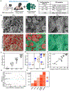
Schematic illustrating a total of 112 patients enrolled in an ongoing, unreported clinical trial (IRB# 10011, Indiana University). From this primary cohort, a blinded subset of 240 SEM images (four technical replicates from each participant) were obtained from n = 60 (randomly selected) participants. A set of 160 SEM images (130 unique) were used to train SEMTWIST for identifying biofilm and nonbiofilm features within SEM images. Following the training phase, the deidentified dataset of 240 images (n = 60) was used to validate SEMTWIST for quantifying biofilm infection abundance in the chronic wound tissue. A random subset (n = 15 of the n = 60) was selected to optimize SEMTWIST’s performance by comparing its results with those from human rater assessments, WGA staining, culture pathology, and PNA-FISH techniques (bottom right), as specified in subsequent figures. PNA-FISH, peptide nucleic acid fluorescence in situ hybridization; SEM, scanning electron microscopy; SEMTWIST, scanning electron microscopy-based Trainable Weka (Waikato Environment for Knowledge Analysis) Intelligent Segmentation Technology; WGA, wheat germ agglutinin.
Figure 2.
Figure 2.
Quantification assessment of biofilm infection (BFI) abundance in human chronic wound edge using scanning electron microscopy (SEM)-machine learning (ML). (A) Schema showing BFI detection and quantification in human chronic wound-edge tissues using SEM image acquisition. BFI quantification was performed using (i) traditional human rater or (ii) novel ML-based approaches; (B) Classification of % BFI abundance obtained using ML and corresponding classification of BFI abundance used by expert human raters: no BFI, low (BFIlo), intermediate (BFIint), or high (BFIhi); (C) raw SEM images (top panel) of human wound-edge tissue samples, BFI abundance was processed and quantified using ML (processed image, bottom panel) approach and classified to BFI abundance. Representative images from BFIlo, BFIint, or BFIhi are shown. (D) Human chronic wound-edge tissue BFI classification using SEM-ML demonstrates the feasibility of the approach for clinical application. The data presented were obtained from human chronic wound edges, with a total of 240 images, unique SEM images quantified (n = 60), including four technical replicates from each wound-edge specimen; (E) a subset of 60 images (n = 15 subjects) was selected from the primary cohort (n = 60) for validation of the quantitative SEM-ML data against a traditional “Expert Rater based” subjective assessment of BFI abundance; this assessment utilized 4 rater expert in biofilm biology; (F) regression analysis showing a positive correlation (p < 0.05), with a correlation coefficient (r = 0.82) between the rater-generated results and the ML-generated data; (G) Bland–Altman plot indicating good agreement between BFI abundance data between human raters and SEM-ML analysis; and (H) correlation plot illustrating interrater variability while a strong correlation between the raters′ mean value and ML quantification.
Figure 3.
Figure 3.
Stepwise implementation of biofilm infection (BFI) detection and quantification using trainable Weka intelligent segmentation technology (TWIST), an ML-based approach. A stepwise procedure of BFI quantitative assessment using TWIST (ML) approach, including (i) adjustment and optimizing of the resolution of the raw image obtained using SEM; (ii) input of the raw image to TWIST for segmentation of BFI (green) and non-BFI elements (red), this process is completed in high magnification of the parent raw image for clear visualization; (iii) toggle overlay allows to visualize and confirm the BFI-positive area is adequately marked; (iv) probability thresholding step provides a map of BFI-infected area (black color pixels) and a quantitative% of BFI abundance.
Figure 4.
Figure 4.
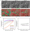
The image pixel density is critical for SEM image biofilm quantification using TWIST. (A) the SEM images were downscaled or upscaled to 320 × 256, 960 × 768, and 2,560 × 2,048 pixels while maintaining the original aspect ratio; (B, C) sensitivity analysis for TWIST performance on five randomly selected SEM images (Supplementary Fig. S3) was performed. The greatest errors occur at lower pixel densities at about 50% (BFIint) biofilm density, (C) images with higher biofilm densities (i.e., >75%, BFIhi) have little or no susceptibility to errors due to pixel density.
Figure 5.
Figure 5.
Training the TWIST to recognize the complex and diverse structure of host tissue in a chronic wound environment. (A) Representative SEM images serving as training set for TWIST to discern some common features in a complex chronic wound-edge tissue depicting noninfected red blood cell (RBC); stomatocytes and echinocytes were observed, fibrin network is dense and classified based on structure (thread-like), blood platelet features, and when activated in a wound environment changes its morphology, which helps in their classification and segmentation, white blood cell (WBC) such as monocytes and neutrophils are enormous in size, mostly embedded within fibrins and extracellular matrix (ECM). Monocytes are observed with pseudo-pod-like projections and blebs, whereas neutrophils have a ruffled morphology. (B) Representative additional images related to ECM used for training the TWIST: ECM images without biofilm infection (top panel) or with biofilm infection (bottom panel). Scale bar: 5,000×.
Figure 6.
Figure 6.
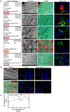
Standard wound culture-based analysis is not reliable to detect fastidious and slow-growing biofilm bacteria in human chronic wounds. (A) Clinical laboratory-based culture pathology reports from four human wound-edge tissue biospecimens out of the cohort of n = 15 showing no microorganism growth indicating negative culture reports. The SEM images with TWIST analysis of the same biospecimen revealed the presence of BFI in these samples. PNA-FISH confirmed the presence of Pseudomonas aeruginosa biofilm (stained red) and Staphylococcus aureus biofilm (stained green) in these samples, scale bar = 5 μm; (B) comparative analysis of wheat germ agglutinin (WGA) BFI staining with SEMTWIST or PNA-FISH assays indicating potential nonspecific detection of BFI with WGA in chronic wound tissues. Top panel: a representative wound biospecimen with no BFI as identified using SEMTWIST; bottom panel: a representative wound biospecimen with BFIhi as identified using SEMTWIST; (C) regression analysis indicating a weak correlation (r = 0.24, p > 0.05) between the WGA-generated and SEMTWIST-generated BFI abundance data.

Similar articles

References

    1. Sen CK, Roy S, Mathew-Steiner SS, et al. Biofilm management in wound care. Plast Reconstr Surg 2021;148(2):275e–288e. - PMC - PubMed
    1. Versey Z, da Cruz Nizer WS, Russell E, et al. Biofilm-innate immune interface: Contribution to chronic wound formation. Front Immunol 2021;12:648554. - PMC - PubMed
    1. Cavallo I, Sivori F, Mastrofrancesco A, et al. Bacterial biofilm in chronic wounds and possible therapeutic approaches. Biology (Basel) 2024;13(2):109. - PMC - PubMed
    1. Bjarnsholt T. The role of bacterial biofilms in chronic infections. APMIS 2013;121(136):1–51. - PubMed
    1. Grooters KE, Ku JC, Richter DM, et al. Strategies for combating antibiotic resistance in bacterial biofilms. Front Cell Infect Microbiol 2024;14:1352273. - PMC - PubMed

LinkOut - more resources