. 2025 Aug 4;222(8):e20241813.
doi: 10.1084/jem.20241813. Epub 2025 May 13.

DNGR-1 regulates proliferation and migration of bone marrow dendritic cell progenitors

Affiliations

DNGR-1 regulates proliferation and migration of bone marrow dendritic cell progenitors

Ana Cardoso et al. J Exp Med. .

Abstract

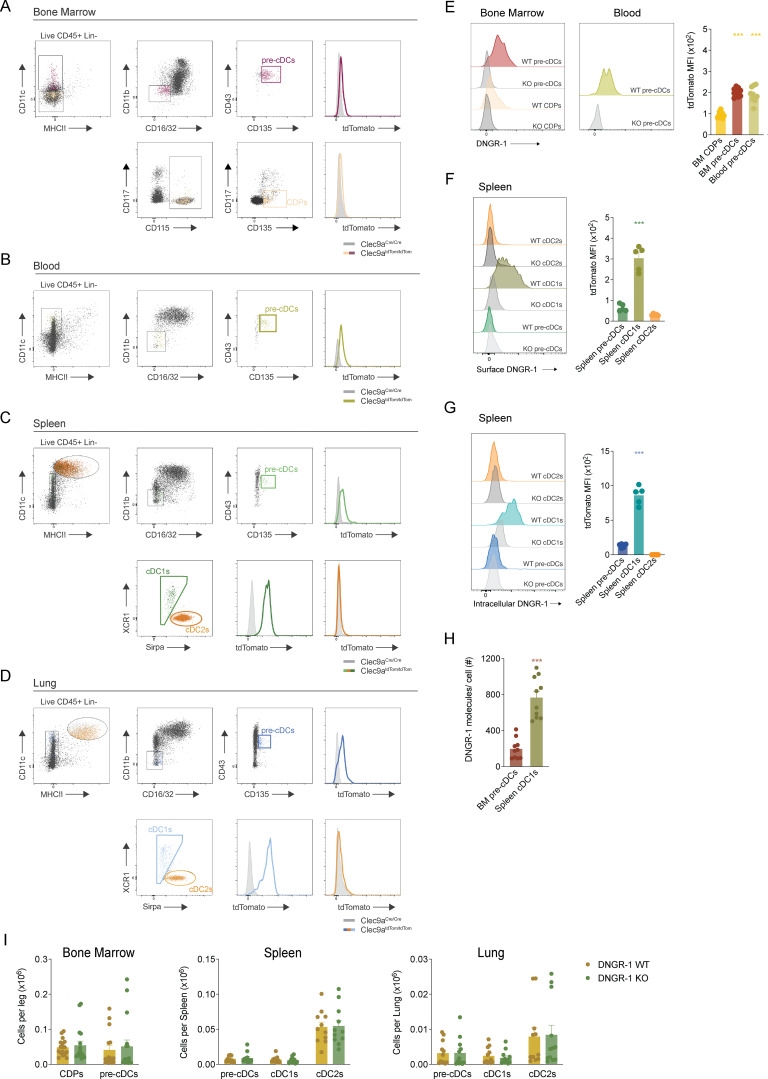
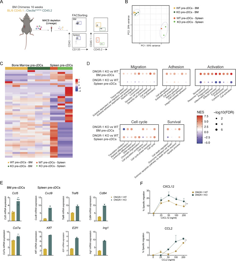
Conventional dendritic cells (cDCs) are sentinel cells that play a crucial role in both innate and adaptive immune responses. cDCs originate from a progenitor (pre-cDC) in the bone marrow (BM) that travels via the blood to seed peripheral tissues before locally differentiating into functional cDC1 and cDC2 cells, as part of a process known as cDCpoiesis. How cDCpoiesis is regulated and whether this affects the output of cDCs is poorly understood. In this study, we show that DNGR-1, an innate immune receptor expressed by cDC progenitors and type 1 cDCs, can regulate cDCpoiesis in mice. In a competitive chimera setting, cDC progenitors lacking DNGR-1 exhibit increased proliferation and tissue migratory potential. Compared with their WT counterparts, DNGR-1-deficient cDC progenitor cells display superior colonization of peripheral tissues but an altered distribution. These findings suggest that cDCpoiesis can be regulated in part by precursor cell-intrinsic processes driven by signals from innate immune receptors such as DNGR-1 that may respond to alterations in the BM milieu.


Conflict of interest statement

Disclosures: K.H.J. Lim reported funding by Wellcome Imperial 4i Clinical Research Fellowship and the Crick Clinical Postdoctoral Fellowship. C. Reis e Sousa reported personal fees from Adendra Therapeutics, Montis Biosciences, and Bicycle Therapeutics outside the submitted work; and additional appointments as Visiting Professor in the Faculty of Medicine at Imperial College London and at King’s College London and an honorary professorship at University College London. No other disclosures were reported.

Figures

Figure 1.
Figure 1.
Low levels of DNGR-1 expression by BM pre-cDCs suffice for mediating XP of dead cell–associated antigens. (A) Uniform manifold approximation and projection (UMAP) analysis of flow cytometry data overlaying different immune populations, namely, effector CD45+ cells overlaid with different cDC lineage populations from BM, spleen, and lung, as identified before (Fig. S1, A–D) in Clec9atdTom/tdTom mice (data are from one mouse, representative of 10). (B) Mean fluorescence intensity (MFI) of tdTom expression in the indicated cell populations of Clec9atdTom/tdTom mice (n = 10). (C) WT BM pre-cDCs and splenic cDC1s were incubated with indicated ratios of DNGR-1 ligand (L)–coated beads to cells. Association of DNGR-1L–coated beads with BM pre-cDCs and splenic cDC1s was measured by flow cytometry. (D and E) (D) WT and DNGR-1 KO BM pre-cDCs or (E) splenic cDC1s were incubated with the indicated ratios of UVC-killed BRAFV600E 5555 cells pulsed with OVA and poly(I:C) or with the indicated concentrations of SIINFEKL. NS represents non-stimulated. Pre-activated OT-I T cells were added and IFN-γ accumulation in culture supernatants was assessed by ELISA 16 h later. Each dot in B represents one mouse, and data are pooled from at least two experiments. Bars represent averages, and error bars represent SEM. Data in C–E depict averages from at least two experiments (total of three to five biological samples; each sample was pooled from two to three mice to achieve one biological replicate); error bars represent SEM. Statistical significance in B was calculated using one-way ANOVA, and in C–E two-way ANOVA with Sidak’s multiple comparisons test. *P < 0.05, **P < 0.01, and ***P < 0.001.
Figure S1.
Figure S1.
FACS analysis of mouse cDCs and their progenitors. (A–D) Gating strategy for CDPs, pre-cDCs, cDC1s, and cDC2s in (A) BM, (B) blood, (C) spleen, and (D) lung. All plots are pre-gated on live (live/dead dye negative) CD45+ lineage (Lin)-negative singlet cells. Lineage cocktail includes CD3, CD19, B220, NK1.1, Ly6G, Ly6D, Siglec-F, and Ter119. (E) Representative plots of anti–DNGR-1 staining (left) in CDPs and pre-cDCs from BM or blood. Right: intensity (MFI) of DNGR-1 staining on the indicated cell populations (n = 10 mice). (F and G) Representative plots of anti–DNGR-1 surface (F) or intracellular (G) labeling in spleen pre-cDCs and cDCs. Right: intensity (MFI) of DNGR-1 staining on the indicated cell populations (n = 5 mice). (H) Quantification of the number of DNGR-1 molecules per cell surface. (I) Quantification of total number of CDPs, pre-cDCs, and cDCs in the BM, spleen, and lung of adult WT (BL/6) and DNGR-1 KO (Clec9aCre/Cre) mice (n = 11–17). Each dot in E, H, and I represents one mouse, and data are pooled from at least two experiments. Data in F and G are from one representative experiment out of three. Bars represent averages, and error bars represent SEM. Statistical significance was calculated in E–G using one-way ANOVA; H was calculated using an unpaired t test with Welch’s correction; and I was calculated using two-way ANOVA with Sidak’s multiple comparisons test. ***P < 0.001. MFI, mean fluorescence intensity.
Figure 2.
Figure 2.
DNGR-1 deficiency subtly affects the kinetics of the cDC network postnatal expansion. (A and B) Quantification of total number of (A) BM CDPs and pre-cDCs and (B) spleen pre-cDCs and cDCs in WT and DNGR-1 KO (Clec9aCre/Cre) at different time points after birth (n = 5–13 mice per time point). (C) Frequency of annexin V+ CDPs, pre-cDCs, and cDCs in BM and/or spleens of WT and DNGR-1 KO (Clec9aeGFP/eGFP) mice at 6 wk of age (n = 12 mice). (D) 6-wk-old mice were injected intraperitoneally with EdU 2 h prior to tissue harvest. Quantification of EdU+ cells in BM and spleen cDC progenitors and spleen cDCs of WT and DNGR-1 KO (Clec9aeGFP/eGFP) mice (n = 10–12 mice). Each dot in A and B represents averages, and data are pooled from three experiments; error bars represent SEM; (C and D) each dot represents one mouse, and data are pooled from two experiments. Bars represent averages and error bars represent SEM. Statistical significance in A and B was calculated using two-way ANOVA with Sidak’s multiple comparisons test, and in C and D was calculated using an unpaired t test with Welch’s correction. *P < 0.05 and **P < 0.01. EdU, 5-ethynyl-2′-deoxyuridine.
Figure 3.
Figure 3.
DNGR-1 KO pre-cDCs are more efficient at peripheral tissue colonization. (A) Lethally irradiated BL/6 CD45.1/2 recipients were reconstituted with a 1:1 ratio of either (a) WT (BL/6 CD45.1) and DNGR-1 KO (Clec9aCre/Cre CD45.2) or (b) WT (BL/6 CD45.1) and WT (BL/6 CD45.2) BM donor cells (2 × 10⁶ total). (B–D) Proportion of CDPs, pre-cDCs, and cDCs of WT:KO or WT:WT origin in (B) BM, (C) spleen, and (D) lung was measured over time (n = 4–5 mice per time point). Each dot in B–D represent averages, and error bars represent SEM. Data are representative from at least one experiment out of three. Statistical significance was calculated using two-way ANOVA with Sidak’s multiple comparisons test. *P < 0.05, **P < 0.01, and ***P < 0.001.
Figure 4.
Figure 4.
DNGR-1 expression impacts gene expression signatures associated with pre-cDC migration. (A) Bulk RNA-seq was performed on BM and splenic pre-cDCs isolated from mixed chimeric mice, with each sample pooled from two mice (n = 6 mice total). (B) Principal component analysis showing clustering of WT or DNGR-1 KO BM and splenic pre-cDCs. (C) Heatmap of the top 50 differentially expressed genes between KO and DNGR-1 WT pre-cDCs derived from BM or spleen. (D) Distribution of the top gene ontology biological processes in KO versus DNGR-1 WT pre-cDCs, in BM and spleen. (E) Differentially expressed genes in WT versus DNGR-1 KO pre-cDCs. (F) BM cells were seeded into the 96-well transwell inserts. After 2 h incubation, cells in the lower well were collected and re-stained. Frequency of BM pre-cDCs recruited toward CXCL12 or CCL2 as assessed by FACS (n = 6 biological replicates). Data are representative from at least one experiment out of three. Each dot in E and F represents a biological replicate; to achieve one biological replicate, cells from two to three mice were pooled. Bars represent averages, and error bars represent SEM. Statistical significance was calculated in E using unpaired t test with Welch’s correction, and in F using two-way ANOVA with Sidak’s multiple comparisons test. *P < 0.05 and **P < 0.01.
Figure S2.
Figure S2.
DNGR-1 KO pre-cDCs are more efficient at peripheral tissues colonization than DNGR-1 WT pre-cDCs. (A) Distribution of the top gene ontology biological processes in KO versus DNGR-1 WT cDC1s in spleen. Data are from the same mice shown in Fig. 4 D. (B) Frequency of BM CDPs and pre-cDCs at week 5 and 24 after transplantation (n = 10–11 mice). Data are from the same mice shown in Fig. 5 B, comparing WT and DNGR-1 KO frequencies over time. (C) Total number of spleen and lung cDC1s and cDC2s pre-cDCs in mixed BM chimeras. Data are from the same mice shown in Fig. 5 B, comparing WT and DNGR-1 KO frequencies over time. Each dot in A represents a biological replicate; to achieve one biological replicate, cells from two mice were pooled. Each dot in B and C represents one mouse, and data are from at least two experiments out of three. Bars represent averages, and error bars represent SEM. Statistical significance was calculated in B and C using two-way ANOVA with Sidak’s multiple comparisons test. ***P < 0.001.
Figure 5.
Figure 5.
DNGR-1 KO pre-cDCs are more efficient at peripheral tissues colonization. (A) Lethally irradiated Clec9aCre/Cre and Clec9aeGFP/eGFP recipients were reconstituted with a 1:1 ratio of Clec9aCre/+R26tdTom/+ (WT) and Clec9aCre/CreR26YFP/YFP (DNGR-1 KO) cells (2 × 10⁶ total donor cells). tdTom and YFP labeling in cDC subsets was normalized to maximum expression of respective donors. (B) Proportion of pre-cDCs and cDCs of WT or DNGR-1 KO origin in spleen (upper panels) and lung (lower panels) at 5 or 24 wk after reconstitution (n = 10–11 mice per time point). (C) Representative images of maximum projection of cryosections (35 µm) from WT:KO mixed chimeras. Spleen (left panels) and lung (right panels) sections (from 5 or 24 wk after reconstitution) were stained with Hoechst and AF647-labeled antibodies (anti–MHC class II, anti-CD64, and anti-B220) (upper). Middle panels show the localization of DNGR-1 WT (tdTom+ YFP−) and of DNGR-1 KO cDC subsets (tdTom− YFP+). (D) Co-localization of WT and DNGR-1 KO pre-cDCs in the spleen and lung at 5 or 24 wk after transplant. (E) Number of WT or DNGR-1 KO pre-cDCs per cluster in the spleen. See Materials and methods for details of analysis. Each dot in B represents one mouse, and data are pooled from at least two experiments out of three. Bars represent averages, and error bars represent SEM. Each dot in D and E corresponds to the analysis of one image from a whole spleen or lung taken from three to seven mice pooled from two experiments shown in B. Statistical significance was calculated in B, D, and E using two-way ANOVA with Sidak’s multiple comparisons test. *P < 0.05, **P < 0.01, and ***P < 0.001.

References

    1. Ahrens, S., Zelenay S., Sancho D., Hanč P., Kjær S., Feest C., Fletcher G., Durkin C., Postigo A., Skehel M., et al. 2012. F-actin is an evolutionarily conserved damage-associated molecular pattern recognized by DNGR-1, a receptor for dead cells. Immunity. 36:635–645. 10.1016/j.immuni.2012.03.008 - DOI - PubMed
    1. Becht, E., McInnes L., Healy J., Dutertre C.A., Kwok I.W.H., Ng L.G., Ginhoux F., and Newell E.W.. 2018. Dimensionality reduction for visualizing single-cell data using UMAP. Nat. Biotechnol. 10.1038/nbt.4314 - DOI - PubMed
    1. Bertheloot, D., Latz E., and Franklin B.S.. 2021. Necroptosis, pyroptosis and apoptosis: An intricate game of cell death. Cell. Mol. Immunol. 18:1106–1121. 10.1038/s41423-020-00630-3 - DOI - PMC - PubMed
    1. Bogunovic, M., Ginhoux F., Helft J., Shang L., Hashimoto D., Greter M., Liu K., Jakubzick C., Ingersoll M.A., Leboeuf M., et al. 2009. Origin of the lamina propria dendritic cell network. Immunity. 31:513–525. 10.1016/j.immuni.2009.08.010 - DOI - PMC - PubMed
    1. Cabeza-Cabrerizo, M., Cardoso A., Minutti C.M., Pereira da Costa M., and Reis e Sousa C.. 2021a. Dendritic cells revisited. Annu. Rev. Immunol. 39:131–166. 10.1146/annurev-immunol-061020-053707 - DOI - PubMed

Substances