. 2025 Jun;18(6):e012350.
doi: 10.1161/CIRCHEARTFAILURE.124.012350. Epub 2025 May 13.

Musclin Counteracts Skeletal Muscle Dysfunction and Exercise Intolerance in Heart Failure With Preserved Ejection Fraction

Affiliations

Musclin Counteracts Skeletal Muscle Dysfunction and Exercise Intolerance in Heart Failure With Preserved Ejection Fraction

Eng Leng Saw et al. Circ Heart Fail. 2025 Jun.

Abstract

Background: Exercise intolerance is a hallmark of heart failure with preserved ejection fraction (HFpEF) and is characterized by skeletal muscle (SkM) dysfunction with impaired oxidative capacity. To maintain oxidative capacity, the SkM secretes myokines such as musclin, which has been shown to potentiate NP (natriuretic peptide) signaling and induce PGC-1α (peroxisome proliferator-activated receptor-γ coactivator-1 alpha) signaling. We sought to investigate the role of musclin in SkM dysfunction in HFpEF. For this study, we selected the oxidative-predominant SkM soleus in HFpEF mice and vastus lateralis from patients with HFpEF.

Methods: Using the SAUNA model, mice underwent HFpEF induction by uninephrectomy, d-aldosterone infusion, and 1% sodium chloride drinking water for 4 weeks. Exogenous musclin was given to HFpEF mice every 2 days during the last 2 weeks of HFpEF induction. Molecular analyses were conducted on blood samples and SkM from HFpEF mice and patients with HFpEF.

Results: In HFpEF mice and patients with HFpEF, increased musclin expression was accompanied by decreased cyclic guanosine monophosphate levels and PGC-1α expression in SkM, suggesting impaired NP signaling. Exogenous administration of musclin in mice with HFpEF demonstrated augmented circulating musclin levels and potentiated NP signaling in SkM as shown by increased PKG1 (protein kinase G1) activity and PGC-1α expression. This was associated with a transition from type-2A to type-1 fiber (type-1 has more endurance) and increased succinate dehydrogenase activity, hindlimb blood flow, and capillary density in the soleus muscle. Exogenous musclin also mitigated cardiac hypertrophy without affecting blood pressure or diastolic function. Most importantly, HFpEF mice treated with musclin demonstrated improved functional and exercise capacity.

Conclusions: Musclin mediates beneficial effects in SkM and heart with improved exercise capacity likely by improving oxidative capacity in SkM. Future studies are warranted to address the therapeutic efficacy of exogenous musclin in humans with HFpEF.

Keywords: exercise tolerance; heart failure; muscle, skeletal; myokines; natriuretic peptides.


Conflict of interest statement

Dr Zamani consulted for Pfizer and Vyaire. Dr Chirinos is an editorial board member of the American Heart Association, the American College of Cardiology, Elsevier, and Wiley. He consulted for Bayer, Fukuda Denshi, Bristol Myers Squibb, JNJ, Edwards Life Sciences, Merck, and NGM Biopharmaceuticals. Dr Sam is a full-time employee of Eli Lilly and Co, Indianapolis, IN. None of the work was funded nor supported by Eli Lilly and Co. The other authors report no conflicts.

Figures
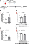
Figure 1.
Figure 1.
Increased musclin and decreased PGC-1α (peroxisome proliferator-activated receptor-γ coactivator-1 alpha) expression in heart failure with preserved ejection fraction (HFpEF) mice. A, HFpEF in C57BL/6J male mice was induced by uninephrectomy, chronic infusion of d-aldosterone, and 1.0% sodium chloride (NaCl) drinking water for 4 weeks. In the soleus muscle of Sham (n=6–7) and HFpEF mice (n=10–11), quantitative analysis showing musclin (Ostn) gene expression (B), musclin protein expression and representative blots (C), PGC-1α (Ppargc1a) gene expression (D), and PGC-1α protein expression and representative blots (E). Data are presented as mean±SEM. Statistical analysis by the Student t test.
Figure 2.
Figure 2.
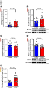
Increased musclin and decreased PGC-1α (peroxisome proliferator-activated receptor-γ coactivator-1 alpha) expression in patients with heart failure with preserved ejection fraction (HFpEF). In the vastus lateralis muscle of control subjects (n=12–13) and patients with HFpEF (n=11–12) from the University of Pennsylvania cohort and quantitative analysis demonstrating musclin (OSTN) gene expression (A), musclin protein expression and representative blots (B), PGC-1α (PPARGC1A) gene expression (C), and PGC-1α protein expression and representative blots (D). Data are presented as mean±SEM. Statistical analysis by the Student t test for normally distributed data (musclin protein expression and PPARGC1A gene expression) or the Mann-Whitney U test for nondistributed data (OSTN gene expression and PGC-1α protein expression). E, Quantitative analysis of circulating musclin levels in control subjects (n=10) and patients with HFpEF (n=20) from the Boston Medical Center cohort. Data are presented as mean±SEM. Statistical analysis by the Student t test.
Figure 3.
Figure 3.
Exogenous musclin activates NPRA (natriuretic peptide receptor-A)/cyclic guanosine monophosphate (cGMP)/PKG1 (protein kinase G1)/PGC-1α (peroxisome proliferator-activated receptor-γ coactivator-1 alpha) signaling in heart failure with preserved ejection fraction (HFpEF) mice. A, Exogenous musclin (0.2 µg/g, every 2 days) was injected intraperitoneally into HFpEF mice in the last 2 weeks of HFpEF induction. B, Illustrative scheme describes musclin’s role in preventing NP (natriuretic peptide) degradation by binding to NPRC (natriuretic peptide clearance receptor) and so further activating NP signaling cascade. In serum samples of Sham (n=11), untreated HFpEF (n=14), and musclin-treated HFpEF mice (n=15), quantitative analysis showing the circulating levels of musclin (C) and BNP (brain natriuretic peptide; D). Data are presented as mean±SEM. Statistical analysis by the Kruskal-Wallis test with the Dunn multiple comparison test. In the soleus muscle of Sham (n=5–6), untreated HFpEF (n=10–11), and musclin-treated HFpEF mice (n=11–12), quantitative analysis showing NPRA (Npr1)-to-NPRC (Npr3) gene expression ratio (E), cGMP levels (F), Pde5a and Pde9a (phosphodiesterase 9A [PDE9]) gene expression (G), VASP (vasodilator-stimulated phosphoprotein) phosphorylation (H), and PGC-1α protein expression with representative blots (I). Data are presented as mean±SEM. Statistical analysis by the 1-way ANOVA with the Tukey multiple comparison test for normally distributed data (cGMP levels, Pde5a and Pde9a gene expression, VASP phosphorylation, and PGC-1α protein expression) or the Kruskal-Wallis test with the Dunn multiple comparison test for nondistributed data (Npr1-to-Npr3 gene expression ratio). 5’-GMP, Pde5a indicates phosphodiesterase 5A (PDE5); and GTP, guanosine triphosphate.
Figure 4.
Figure 4.
NPRA (natriuretic peptide receptor-A)/cyclic guanosine monophosphate (cGMP) signaling in patients with heart failure with preserved ejection fraction (HFpEF). In the vastus lateralis muscle of control subjects and patients with HFpEF from the University of Pennsylvania cohort, quantitative analysis showing NPRA (NPR1)-to-NPR3 (natriuretic peptide receptor 3 [NPRC]) gene expression ratio (n=9–11/group; A), cGMP levels (n=7–8/group; B), and PDE5A and PDE9A gene expression (n=11–13/group; C). Data are presented as mean±SEM. Statistical analysis by the Student t test for normally distributed data (PDE5A and PDE9A gene expression) or the Mann-Whitney U test for nondistributed data (NPR1-to-NPR3 gene expression ratio and cGMP levels).
Figure 5.
Figure 5.
Exogenous musclin enhances fiber transition and SDH (succinate dehydrogenase) activity in heart failure with preserved ejection fraction (HFpEF) mice. A, Quantitative analysis showing the abundance of type-1/2A hybrid fibers in the soleus muscle of Sham (n=5), untreated HFpEF (n=4), and musclin-treated HFpEF mice (n=5). Data are presented as mean±SEM. Statistical analysis by the Kruskal-Wallis test with the Dunn multiple comparison test. B, Representative confocal images showing staining of dystrophin (white), MHCI (myosin heavy chain I) in type-1 fiber (green), MHCIIA (myosin heavy chain IIA) in type-2A fiber (red), and MHCIIX (myosin heavy chain IIX) in type-2X fiber (blue) in the soleus muscle. Type-1/2A hybrid fibers (denoted by the yellow arrows) are coexpressing MHCI (green) and MHCIIA (red). C, Qualitative immunoblot and quantitative analysis showing MHCI and MHCIIA protein expression and representative blots in the soleus muscle of Sham (n=4–5), untreated HFpEF (n=11), and musclin-treated HFpEF mice (n=11–12). Data are presented as mean±SEM. Statistical analysis by the 1-way ANOVA with the Tukey multiple comparison test. D, Quantitative analysis showing the SDH activity in the soleus muscle of Sham (n=5), untreated HFpEF (n=5), and musclin-treated HFpEF mice (n=5). Data are presented as mean±SEM. Statistical analysis by the 1-way ANOVA with the Tukey multiple comparison test. E, Representative brightfield images showing the SDH activity in the soleus muscle.
Figure 6.
Figure 6.
Exogenous musclin increases hindlimb blood flow and capillary density in heart failure with preserved ejection fraction (HFpEF) mice. A, Quantitative analysis and showing the blood flow in the hindlimbs of Sham (n=10), untreated HFpEF (n=16), and musclin-treated HFpEF mice (n=17). Data are presented as mean±SEM. Statistical analysis by the 1-way ANOVA with the Tukey multiple comparison test. B, Representative laser Doppler images showing blood flow in the hindlimbs of mice. C, Quantitative analysis showing the density of capillaries in the soleus muscle of Sham (n=5), untreated HFpEF (n=5), and musclin-treated HFpEF mice (n=5). Data are presented as mean±SEM. Statistical analysis by the 1-way ANOVA with the Tukey multiple comparison test. D, Representative confocal images showing staining of dystrophin (white; white asterisk indicates a single fiber), isolectin-stained endothelial cells (red; denoted by the yellow arrows), and nuclei (blue) in the soleus muscle. E, Quantitative analysis showing the relative gene expression of Pdgfb and Angpt2 in the soleus muscle of Sham (n=5), untreated HFpEF (n=9–10), and musclin-treated HFpEF mice (n=11–12). Data are presented as mean±SEM. Statistical analysis by the 1-way ANOVA with the Tukey multiple comparison test.
Figure 7.
Figure 7.
Exogenous musclin reduces cardiomyocyte hypertrophy and Nppb gene expression in the left ventricle (LV) of heart failure with preserved ejection fraction (HFpEF) mice. A, Quantitative analysis showing the cardiomyocyte size in the LV of Sham (n=6), untreated HFpEF (n=10), and musclin-treated mice (n=8). Data are presented as mean±SEM. Statistical analysis by the 1-way ANOVA with the Tukey multiple comparison test. B, Representative brightfield images of hematoxylin and eosin-stained LV sections. C, Quantitative analysis showing the relative gene expression of Nppb in the LV of Sham (n=10), untreated HFpEF (n=10), and musclin-treated HFpEF mice (n=10). Data are presented as mean±SEM. Statistical analysis by the 1-way ANOVA with the Tukey multiple comparison test. D, Quantitative analysis showing the fibrotic area in the LV of Sham (n=6), untreated HFpEF (n=11), and musclin-treated mice (n=9). Data are presented as mean±SEM. Statistical analysis by the 1-way ANOVA with the Tukey multiple comparison test. E, Representative brightfield images of picrosirius red–stained LV sections.
Figure 8.
Figure 8.
Exogenous musclin improves treadmill exercise exhaustion capacity in heart failure with preserved ejection fraction (HFpEF) mice. A quantitative analysis demonstrating treadmill running distance (A) and running time (B) of untreated HFpEF and musclin-treated HFpEF mice (n=10–11/group). Data are presented as mean±SEM. Statistical analysis by the Mann-Whitney U test.

Comment in

References

    1. Solomon SD, McMurray JJV, Claggett B, de Boer RA, DeMets D, Hernandez AF, Inzucchi SE, Kosiborod MN, Lam CSP, Martinez F, et al. ; DELIVER Trial Committees and Investigators. Dapagliflozin in heart failure with mildly reduced or preserved ejection fraction. N Engl J Med. 2022;387:1089–1098. doi: 10.1056/NEJMoa2206286 - PubMed
    1. Kosiborod MN, Abildstrøm SZ, Borlaug BA, Butler J, Rasmussen S, Davies M, Hovingh GK, Kitzman DW, Lindegaard ML, Møller DV, et al. ; STEP-HFpEF Trial Committees and Investigators. Semaglutide in patients with heart failure with preserved ejection fraction and obesity. N Engl J Med. 2023;389:1069–1084. doi: 10.1056/NEJMoa2306963 - PubMed
    1. Roh J, Hill JA, Singh A, Valero-Muñoz M, Sam F. Heart failure with preserved ejection fraction: Heterogeneous syndrome, diverse preclinical models. Circ Res. 2022;130:1906–1925. doi: 10.1161/CIRCRESAHA.122.320257 - PMC - PubMed
    1. Pandey A, Shah SJ, Butler J, Kellogg DL, Jr, Lewis GD, Forman DE, Mentz RJ, Borlaug BA, Simon MA, Chirinos JA, et al. Exercise intolerance in older adults with heart failure with preserved ejection fraction: JACC state-of-the-art review. J Am Coll Cardiol. 2021;78:1166–1187. doi: 10.1016/j.jacc.2021.07.014 - PMC - PubMed
    1. Saw EL, Ramachandran S, Valero-Muñoz M, Sam F. Skeletal muscle (dys)function in heart failure with preserved ejection fraction. Curr Opin Cardiol. 2021;36:219–226. doi: 10.1097/HCO.0000000000000824 - PMC - PubMed

MeSH terms

Substances