. 2025 May 13;167(1):137.
doi: 10.1007/s00701-025-06555-y.

Tumor resection in paramedian structures of the frontal lobe poses a risk for corpus callosum infarction

Affiliations

Tumor resection in paramedian structures of the frontal lobe poses a risk for corpus callosum infarction

Yoshiteru Shimoda et al. Acta Neurochir (Wien). .

Abstract

Purpose: Surgeons resecting intraparenchymal tumors should be aware of potential white matter ischemia resulting from damage to the medullary artery arising from the cerebral cortex. In the vicinity of the paramedian structure, crucial brain regions for higher brain function such as corpus callosum and cingulate cortex are located. However, the actual area of ischemia induced by damaging the medullary artery supplying the paramedian structures is not known. The present study investigated the ischemic field following tumor resection in paramedian structures of the frontal lobe.

Methods: Patients having intraparenchymal tumors with lesions in the paramedian structures of the frontal lobe (superior frontal gyrus or cingulate gyrus) resected between April 2016 and June 2022 at Tohoku University Hospital were included in the study. Magnetic resonance images obtained within 72 h after surgery were used to retrospectively examine the extent of the resection and the distribution of ischemic complications. Related postoperative clinical symptoms were assessed using medical records.

Results: Thirty-three cases matched the inclusion criteria. The median age was 48 years. Cases comprised patients with an astrocytoma IDH-mutant (n = 11), oligodendroglioma IDH-mutant, and 1p/19q-codeletion (n = 12), and glioblastoma IDH-wildtype (n = 10). The main locations were superior frontal gyrus only (n = 17), cingulate gyrus only (n = 8), and both the frontal lobe and cingulate gyrus (n = 8). The cingulate gyrus was removed in 19 cases. In 16 of the 19 cases, ischemic foci were observed in the adjacent corpus callosum. In the 14 cases in which the cingulate gyrus was not removed, no ischemic foci appeared in the corpus callosum. Three cases exhibited a prolonged disturbance of consciousness after the second postoperative day, all with corpus callosum infarction.

Conclusion: Surgeons resecting intraparenchymal tumors in the paramedian structures of the frontal lobe, especially the cingulate gyrus, should be aware of the potential for ischemia foci emerging in the corpus callosum.

Keywords: Cingulate gyrus; Corpus callosum; Disconnection syndrome; Surgery.


Conflict of interest statement

Declarations. Ethics approval: This was approved by the Institutional Review Board Committee of Tohoku University Hospital (2023–1-416). Consent to participate and to publish: NA (Participants were given the option to opt out of this study). Conflict of interest: The authors declare no competing interests.

Figures

Fig. 1
Fig. 1
Typical ischemic lesion after cingulate gyrus resection without injury to the corpus callosum. A 24-year-old woman underwent MRI for evaluation of headaches. An enlarged T2 high-signal area was observed in the anterior cingulate gyrus (A). A low-grade glioma was suspected, and total resection was performed without injury to the corpus callosum (B). Postoperative MRI showed an ischemic area in the corpus callosum adjacent to the resected area (C). Postoperatively, the patient exhibited no new neurologic symptoms
Fig. 2
Fig. 2
Cingulate gyrus resection results in corpus callosum infarction. The cingulate gyrus was removed in 19 cases. In 8 of these cases, the corpus callosum was also partially resected. In these 8 cases, 5 exhibited ischemia of the corpus callosum (63%). In the 3 cases with corpus callosum ischemia, the corpus callosum directly below the resected cingulate gyrus was also removed, making it difficult to evaluate ischemic lesions of the corpus callosum. In 11 cases, the cingulate gyrus was removed but the corpus callosum was completely preserved. In all 11 cases, ischemia was observed at the lateral side of the corpus callosum (100%). In contrast, in 14 cases in which the cingulate gyrus was not resected, no ischemia of the corpus callosum was observed (0%)
Fig. 3
Fig. 3
Extent of the ischemic region in the corpus callosum plotted coaxially with the extent of the cingulate gyrus resection
Fig. 4
Fig. 4
A 67-year-old man underwent an incidental MRI. The cingulate gyrus was enlarged, extended along the long axis, and exhibited a high T2 signal (A). A low-grade glioma was suspected and resection was performed. The entire high T2 signal area was removed (B). The resection extended 5 cm longitudinally (C). Postoperative MRI showed an ischemic area in the corpus callosum lateral to the resected region (D). Postoperatively, he exhibited consciousness disturbances for 7 days
Fig. 5
Fig. 5
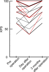
The temporal changes in Karnofsky performance scale (KPS) in the group of cases with no lesions in the cingulate gyrus that caused no corpus callosum infarction (black line) and the group of cases with lesions in the cingulate gyrus that caused corpus callosum infarction due to cingulate gyrus removal (red line), before and at day after surgery, and at 3 months after surgery. The data for the 2 patients who had had a relapse at 3 months, were excluded. In both groups, KPS temporarily decreased after surgery, but tended to recover by 3 months after surgery. There was no difference in the degree of recovery between the two groups (p = 0.19)
Fig. 6
Fig. 6
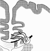
Microvascular images of the corpus callosum obtained from the literature. Modified from T Okudera et al. Neuropathology 1999[9]. Note that the perforating artery supplying the outer part of the corpus callosum runs through the cingulate gyrus (arrow), suggesting that infarction of the corpus callosum is inevitable when the cingulate gyrus is resected
Fig. 7
Fig. 7
The combination of intra-arterial administration of contrast medium and ultrahigh-resolution computed tomography angiography allowed for observation of the perforating artery supplying the outer part of the corpus callosum running through the cingulate gyrus (arrow). The 3D tractography of the corpus callosum, derived from diffusion tensor imaging, is being fused

References

    1. Akashi T, Takahashi S, Mugikura S, Sato S, Murata T, Umetsu A, Takase K (2017) Ischemic white matter lesions associated with medullary arteries: classification of mri findings based on the anatomic arterial distributions. AJR Am J Roentgenol 209(3):W160–W168 - PubMed
    1. Degos JD, Gray F, Louarn F, Ansquer JC, Poirier J, Barbizet J (1987) Posterior callosal infarction. Clinicopathological correlations. Brain 110 ( Pt 5)(5):1155–1171 - PubMed
    1. Gomes FB, Dujovny M, Umansky F, Berman SK, Diaz FG, Ausman JI, Mirchandani HG, Ray WJ (1986) Microanatomy of the anterior cerebral artery. Surg Neurol 26(2):129–141 - PubMed
    1. Graham D, Tisdall MM, Gill D (2016) Corpus callosotomy outcomes in pediatric patients: A systematic review. Epilepsia 57(7):1053–1068 - PubMed
    1. Kahilogullari G, Comert A, Arslan M, Esmer AF, Tuccar E, Elhan A, Tubbs RS, Ugur HC (2008) Callosal branches of the anterior cerebral artery: an anatomical report. Clin Anat 21(5):383–388 - PubMed

MeSH terms

LinkOut - more resources