Comparing the outcome after double level osteotomies in severe valgus and varus knees
- PMID: 40358751
- PMCID: PMC12075348
- DOI: 10.1007/s00402-025-05893-x
Comparing the outcome after double level osteotomies in severe valgus and varus knees
Abstract
Introduction: Osteotomies have played an important role in joint preservation surgery of the knee joint for many years. A double level osteotomy is performed for severe varus or valgus deformities. There are numerous publications on double level osteotomies for severe varus deformities, whereas there are no publications on valgus deformities. The hypothesis of this study was to compare the clinical outcome after varus DLO with that after valgus DLO.
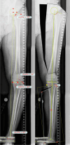
Material and methods: In this retrospective study, 40 DLOs were followed up in 34 patients. In group one (13 cases, age 45.6 (16-61) years) a varization DLO was performed, in group two (24 cases, age 48.3 (20-61) years) a valgization DLO was performed. The pre- and postoperative clinical scores were recorded: Tegner Activity score, Japanese knee society Score and Lysholm Score. The leg axis and knee joint angles were recorded and compared pre- and postoperatively.
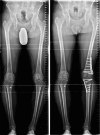
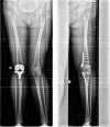
Results: The follow-up period was 24 (6-81) months. The follow-up rate was 73% (27/37). The preoperative leg axis in group one showed an average valgus of 15.9° (9-40°). Group two had an average varus of 12° (8-21°). Postoperatively, the leg axis was 3.4° varus in group one and 0.5° valgus in group two. The mLDFA changed in group one from 83.2° to 90.9°, the MPTA from 95.5° to 87.0°. In group two, the mLDFA changed from 91.9° to 85.9° and the MPTA from 83.3° to 88.3° on average. The JLCA changed in group one from - 3.2 (- 5°-0°) to - 0.5° (- 3-2°) postoperative and in group two from 3.3° (1-8°) to 3.0° (0-6°) postoperative. Tegner score, Lysholm score and Japanese knee Society score all improved significantly in both groups. Patients with a valgus axis have worse clinical scores before surgery than the varus group, but the varus group shows a higher potential for improvement postoperatively. Every patient stated that they would have the operation performed again. Complications were rare, two overcorrections required corrective surgery. Two hinge fractures were treated intraoperatively with additional contralateral plate osteosynthesis.
Conclusions: Patients show very good clinical results after DLO. The improvements in the valgus knees are greater, but starting from a lower preoperative level, probably due to improvements in both the lateral compartment and the patellofemoral compartment. An important finding was that JLCA is normalizing in valgization DLO but not in varization DLO. This needs to be considered in planning a DLO.
Keywords: DLO; Double level osteotomy; Genu valgum; Genu varum; Valgization; Varization.
© 2025. The Author(s).
Conflict of interest statement
Declarations. Conflict of interest: The authors declare no competing interests.
Figures


References
Publication types
MeSH terms
LinkOut - more resources
Full Text Sources

