. 2025 May 13;135(13):e188541.
doi: 10.1172/JCI188541. eCollection 2025 Jul 1.

Activation of intestinal endogenous retroviruses by alcohol exacerbates liver disease

Affiliations

Activation of intestinal endogenous retroviruses by alcohol exacerbates liver disease

Noemí Cabré et al. J Clin Invest. .

Abstract

Alcohol-associated liver disease represents a significant global health challenge, with gut microbial dysbiosis and bacterial translocation playing a critical role in its pathogenesis. Patients with alcohol-associated hepatitis had increased fecal abundance of mammalian viruses, including retroviruses. This study investigated the role of endogenous retroviruses (ERVs) in the development of alcohol-associated liver disease. Transcriptomic analysis of duodenal and liver biopsies revealed increased expression of several human ERVs, including HERV-K and HERV-H, in patients with alcohol-associated liver disease compared with individuals acting as controls. Chronic-binge ethanol feeding markedly induced ERV abundance in intestinal epithelial cells but not the livers of mice. Ethanol increased ERV expression and activated the Z-DNA binding protein 1 (Zbp1)-mixed lineage kinase domain-like pseudokinase (Mlkl) signaling pathways to induce necroptosis in intestinal epithelial cells. Antiretroviral treatment reduced ethanol-induced intestinal ERV expression, stabilized the gut barrier, and decreased liver disease in microbiota-humanized mice. Furthermore, mice with an intestine-specific deletion of Zbp1 were protected against bacterial translocation and ethanol-induced steatohepatitis. These findings indicate that ethanol exploits this pathway by inducing ERVs and promoting innate immune responses, which results in the death of intestinal epithelial cells, gut barrier dysfunction, and liver disease. Targeting the ERV/Zbp1 pathway may offer new therapies for patients with alcohol-associated liver disease.

Keywords: Gastroenterology; Hepatology; Microbiome; Molecular biology.


Figures
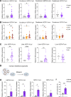
Figure 1
Figure 1. Upregulation of human endogenous retroviruses in duodenal biopsies of patients with alcohol-associated liver disease.
(A) Duodenal biopsies were obtained from individuals without alcohol use disorder or alcohol-associated liver disease (controls; n = 16) and patients with alcohol use disorder and alcohol-associated liver disease (n = 44), and qPCR was performed to measure mRNA expression of human endogenous retroviruses (HERVs). (B) Duodenal expression levels of HERVs in patients with advanced fibrosis (F2–F4) compared with individuals acting as controls and patients with no or mild liver disease (F0–F1). (C) Liver biopsies were obtained from individuals without alcohol use disorder or alcohol-associated liver disease (controls, n = 5) and patients with alcohol use disorder and alcohol-associated liver disease (n = 27), and qPCR was performed to measure mRNA expression of HERVs. (D) Schematic representation of the experiment. (DF) Human intestinal organoids were treated with ethanol (50 mM) for 24 hours to assess HERV expression (E) and to measure cytotoxicity using the lactate dehydrogenase (LDH) assay of human organoids (F). (E and F) Results were generated from 2 technical replicates. P values among groups were determined by Mann-Whitney U test (A, C, E, and F) or 1-way ANOVA with 2-stage step-up method of Benjamini, Krieger and Yekutieli test (B). Results are expressed as mean ± SEM. *P < 0.05, **P < 0.01, ****P < 0.0001.
Figure 2
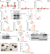
Figure 2. Ethanol activates Zbp1 and induces Mlkl-mediated necroptosis in intestinal epithelial cells.
(A) Germ-free C57BL/6 mice were colonized with fecal microbiota from patients with alcohol-associated hepatitis (AH) and fed oral isocaloric (control) or chronic-binge ethanol diets. (B) Intestinal and hepatic levels of mouse mammary tumor virus (MMTV) env and gag mRNA. (C) Level of Zbp1 mRNA in the ileum. (D) Intestinal epithelial cells were isolated from ethanol-fed mice, and immunoblots were performed for phospho-Mlkl, Mlkl, Zbp1, and Gapdh; protein amounts of Zbp1 relative to Gapdh and protein amounts of phospho-Mlkl relative to Mlkl are shown (n = 5 control, n = 6 ethanol). (E–J) Mouse intestinal organoids were incubated with ethanol (0, 10, 20, and 50 mM) for 24 hours. (E) Expression levels of MMTV env and gag mRNA. (F) Expression level of Zbp1 mRNA. (G) Immunoblots of phospho-Mlkl, Mlkl, and Gapdh; protein amounts of phospho-Mlkl relative to Mlkl are shown (n = 5 in each group). (H) Immunoblot analysis of Mlkl in cytoplasmic and membrane extracts. Nonreducing and reducing SDS-PAGE were used to detect oligomerized and total Mlkl forms, respectively. (I) Representative images of intestinal organoids before and after treatment with ethanol (50 mM). (J) Lactate dehydrogenase (LDH) assay of supernatants was performed to measure cytotoxicity. Results were generated from 3 (B) or 2 (C–J) technical replicates. P values among groups were determined by Mann-Whitney U test (B–D) or 1-way ANOVA with Tukey’s post hoc test (E–G and J). Results are expressed as mean ± SEM. FMT, fecal microbiota transplantation. *P < 0.05, **P < 0.01, ***P < 0.001, ****P < 0.0001. AU, arbitrary units.
Figure 3
Figure 3. Ethanol-induced necroptosis is reduced by suppressing ERV expression or inhibiting Mlkl in intestinal organoids.
(A) Mouse intestinal organoids were treated with the antiretroviral agents emtricitabine (EC; 100 μM) or tenofovir disoproxil fumarate (TDF; 100 μM) and incubated with ethanol (50 mM) for 24 hours. (B) Expression levels of mouse mammary tumor virus (MMTV) env and gag mRNA. (C) Expression level of Zbp1 mRNA. (D) Immunoblots of phospho-Mlkl, Mlkl, and Gapdh; protein amounts of phospho-Mlkl relative to Mlkl are shown (n = 4 in each group). (E) Lactate dehydrogenase (LDH) assay of supernatants was performed to measure cytotoxicity. (F) Mouse intestinal organoids were treated with the Mlkl inhibitor GW806742X (2 μM) and incubated with ethanol (50 mM) for 24 hours. (G) Expression levels of MMTV env and gag mRNA. (H) Expression level of Zbp1 mRNA. (I) Immunoblots of phospho-Mlkl, Mlkl, and Gapdh; protein amounts of phospho-Mlkl relative to Mlkl are shown (n = 5 in each group). (J) Lactate dehydrogenase assay of supernatant was performed to measure cytotoxicity. Results were generated from 3 (B and C) or 2 (DJ) technical replicates. P values among groups were determined by 1-way ANOVA with 2-stage step-up method of Benjamini, Krieger and Yekutieli test (B–E) or Tukey’s post hoc test (G–J). Results are expressed as mean ± SEM. RTi, reverse transcriptase inhibitor. *P < 0.05, **P < 0.01, ***P < 0.001, ****P < 0.0001. AU, arbitrary units.
Figure 4
Figure 4. Overexpression of HERV-K induces necroptosis in ethanol-treated intestinal cells.
(A) MODE-K cells were infected with a lentiviral control vector (Mock) or a lentivirus overexpressing human endogenous retroviruses K (HERV-K). Infected MODE-K cells were treated with 50 mM ethanol and the Mlkl inhibitor GW806742X (2 μM) for 24 hours. (B) Expression levels of HERV-K mRNA. (C) Expression level of Zbp1 mRNA. (D) Lactate dehydrogenase (LDH) assay of supernatant was performed to measure cytotoxicity. (B–D) Results were generated from 4 technical replicates. P values among groups were determined by 1-way ANOVA with Tukey’s post hoc test. Results are expressed as mean ± SEM. *P < 0.05, **P < 0.01, ****P < 0.0001.
Figure 5
Figure 5. Antiretroviral treatment reduces ethanol-induced intestinal necroptosis and liver disease.
(A) Germ-free C57BL/6 mice were colonized with fecal microbiota from patients with alcohol-associated hepatitis (AH) and treated with a combination of antiretrovirals (660 μM emtricitabine, 314 μM tenofovir, and 375 μM nevirapine) in the drinking water after second colonization and then in liquid diet. Gnotobiotic mice were fed oral isocaloric (control) or chronic-binge ethanol diets. (B) Levels of mouse mammary tumor virus (MMTV) env and gag mRNA in the ileum. (C) Levels of MMTV env and gag mRNA in intestinal epithelial cells (IECs) isolated from the small intestine. (D) Levels of Zbp1 mRNA in the ileum and in intestinal epithelial cells isolated from the small intestine. (E and F) Intestinal epithelial cells were isolated from ethanol-fed mice, and immunoblots were performed for phospho-Mlkl, Mlkl, Zbp1, and Gapdh; protein amounts of Zbp1 relative to Gapdh and protein amounts of phospho-Mlkl relative to Mlkl are shown (n = 8 in each group). (G) Serum levels of ALT. (H) Hepatic triglyceride content. (I) Representative images of liver sections stained with Oil Red O (scale bars: 200 μm) and quantification of Oil Red O-stained area. (J) Hepatic levels of Cxcl1 and Cxcl2 mRNA. (K) Colony-forming units on MacConkey agar plates from the liver. (L) Hepatic E. coli abundance normalized to bacterial 16S, as measured by qPCR. (B–L) Results were generated from 2 technical replicates. P values among groups were determined by 1-way ANOVA with Tukey’s post hoc test (B; D, left; G–I; and L) or Mann-Whitney U test (C, D, right, F, J, and K). Results are expressed as mean ± SEM. *P < 0.05, **P < 0.01, ****P < 0.0001. AU, arbitrary units.
Figure 6
Figure 6. Deficiency of Zbp1 in intestinal epithelial cells attenuates ethanol-induced intestinal necroptosis and liver disease.
Mice lacking Zbp1 in intestinal epithelial cells (Zbp1ΔIEC) and their Zbp1fl/fl littermate controls were fed oral isocaloric (control) or chronic-binge ethanol diets. (A) Serum levels of ALT. (B) Hepatic triglyceride content. (C) Representative images of liver sections stained with Oil Red O (scale bars: 200 μm) quantification of Oil Red O–stained area. (D) Hepatic levels of Ccl2 and Cxcl2 mRNA. (E) Colony-forming units on MacConkey agar plates from the liver. (F) Hepatic E. coli abundance normalized to bacterial 16S, as measured by qPCR. (G) Levels of MMTV env and gag mRNA in the ileum. (H) Immunoblots of phospho-Mlkl, Mlkl, and Gapdh; protein amounts of phospho-Mlkl relative to Mlkl are shown (n = 5 in each group). Results were generated from 2 technical replicates. P values among groups were determined by 1-way ANOVA with Tukey’s post hoc test (A–D, F, and G) or Mann-Whitney U test (E and H). Results are expressed as mean ± SEM. *P < 0.05, **P < 0.01, ****P < 0.0001.*P < 0.05, **P < 0.01, ***P < 0.001, ****P < 0.0001. AU, arbitrary units.

References

    1. Aberg F, et al. Alcohol-associated liver disease-global epidemiology. Hepatology. 2024;80(6):1307–1322. doi: 10.1097/HEP.0000000000000899. - DOI - PubMed
    1. Narro GEC, et al. Alcohol-related liver disease: a global perspective. Ann Hepatol. 2024;29(5):101499. doi: 10.1016/j.aohep.2024.101499. - DOI - PubMed
    1. Hsu CL, Schnabl B. The gut-liver axis and gut microbiota in health and liver disease. Nat Rev Microbiol. 2023;21(11):719–733. doi: 10.1038/s41579-023-00904-3. - DOI - PMC - PubMed
    1. Fairfield B, Schnabl B. Gut dysbiosis as a driver in alcohol-induced liver injury. JHEP Rep. 2021;3(2):100220. doi: 10.1016/j.jhepr.2020.100220. - DOI - PMC - PubMed
    1. Wang L, et al. Intestinal REG3 lectins protect against alcoholic steatohepatitis by reducing mucosa-associated microbiota and preventing bacterial translocation. Cell Host Microbe. 2016;19(2):227–239. doi: 10.1016/j.chom.2016.01.003. - DOI - PMC - PubMed