. 2025 May 13;135(13):e180826.
doi: 10.1172/JCI180826. eCollection 2025 Jul 1.

The gut microbiome controls reactive astrocytosis during Aβ amyloidosis via propionate-mediated regulation of IL-17

Affiliations

The gut microbiome controls reactive astrocytosis during Aβ amyloidosis via propionate-mediated regulation of IL-17

Sidhanth Chandra et al. J Clin Invest. .

Abstract

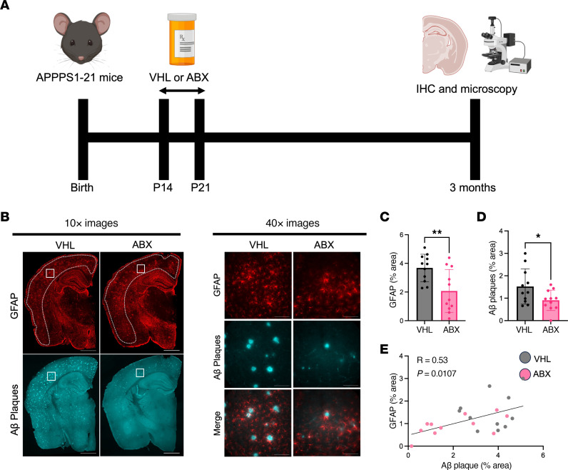
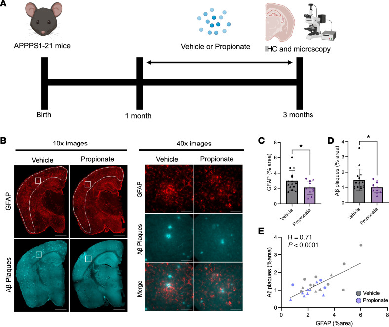
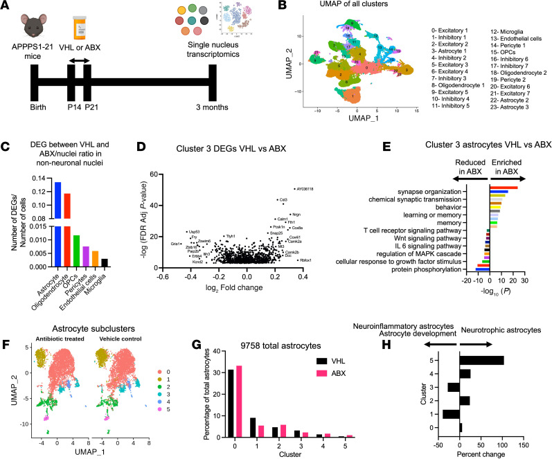
Accumulating evidence implicates the gut microbiome (GMB) in the pathogenesis and progression of Alzheimer's disease (AD). We recently showed that the GMB regulates reactive astrocytosis and Aβ plaque accumulation in a male APPPS1-21 AD mouse model. Yet, the mechanism(s) by which GMB perturbation alters reactive astrocytosis in a manner that reduces Aβ deposition remain unknown. Here, we performed metabolomics on plasma from mice treated with antibiotics (ABX) and identified a significant increase in plasma propionate, a gut-derived short-chain fatty acid, only in male mice. Administration of sodium propionate reduced reactive astrocytosis and Aβ plaques in APPPS1-21 mice, phenocopying the ABX-induced phenotype. Astrocyte-specific RNA-Seq on ABX- and propionate-treated mice showed reduced expression of proinflammatory and increased expression of neurotrophic genes. Next, we performed flow cytometry experiments, in which we found that ABX and propionate decreased peripheral RAR-related orphan receptor-γ+ (Rorγt+) CD4+ (Th17) cells and IL-17 secretion, which positively correlated with reactive astrocytosis. Last, using an IL-17 mAb to deplete IL-17, we found that propionate reduced reactive astrocytosis and Aβ plaques in an IL-17-dependent manner. Together, these results suggest that gut-derived propionate regulates reactive astrocytosis and Aβ amyloidosis by decreasing peripheral Th17 cells and IL-17 release. Thus, propionate treatment or strategies boosting propionate production may represent novel therapeutic strategies for the treatment of AD.

Keywords: Alzheimer disease; Cellular immune response; Immunology; Microbiology; Neuroscience.


Conflict of interest statement

Conflict of interest: The authors have declared that no conflict of interest exists.

Figures

Figure 1
Figure 1. ABX-mediated GBM perturbation reduces GFAP+ astrocytes and Aβ plaques in the cortex of APPPS1-21 male mice.
(A) Schematic depicting the experimental paradigm. (B) Representative images of whole brain sections (original magnification, ×10) and high-magnification images of cortex (original magnification, ×40) stained for GFAP+ astrocytes and Aβ plaques in APPPS1-21 male mice treated with ABX or VHL control. Quantification of the cortical (C) GFAP+ astrocyte percentage area and (D) the Aβ plaque percentage area in VHL- and ABX-treated APPPS1-21 mice. (E) Pearson’s correlation analysis between GFAP+ astrocyte percentage area and Aβ plaque percentage area in VHL- and ABX-treated APPPS1-21 mice. Data are expressed as the mean ± SD. n = 11/group. Statistics were calculated using a 2-tailed, unpaired Student’s t test. n = 4 sections per animal. *P ≤ 0.05 and **P ≤ 0.01. Scale bars: 1,000 μm (×10 images) and 100 μm (×40 images). Dotted lines indicate the analyzed area of the cortex.
Figure 2
Figure 2. ABX-mediated GBM perturbation increases plasma propionate levels, which negatively correlate with reactive astrocytosis in APPPS1-21 male mice.
(A) Schematic depicting the experimental paradigm. Relative GC-MS quantification of the SCFAs (B) propionate, (C) acetate, and (D) butyrate in the plasma of VHL- and ABX-treated APPPS1-21 male (M) and female (F) mice. Pearson’s correlation analysis between (E) GFAP astrocyte percentage area and (F) C3 area/GFAP area and plasma propionate levels in VHL- and ABX-treated male APPPS1-21 mice. Data are expressed as the mean ± SD. n = 6–7/group. Statistics were calculated using 2-way ANOVA. *P ≤ 0.05 and **P ≤ 0.01.
Figure 3
Figure 3. Exogenous propionate treatment reduces GFAP+ astrocytes and Aβ plaques in male and female APPPS1-21 mice.
(A) Schematic depicting the experimental paradigm. (B) Representative images of whole brain sections (original magnification, ×10) and cortex (original magnification, ×40) tissue stained for GFAP+ astrocytes and Aβ plaques in APPPS1-21 mice treated with VHL control or propionate. Quantification of cortical (C) GFAP+ astrocyte percentage area and (D) Aβ plaque percentage area in VHL- and propionate-treated male and female APPPS1-21 mice. (E) Pearson’s correlation analysis between GFAP+ astrocyte percentage area and Aβ plaque percentage area in VHL- and propionate-treated male and female APPPS1-21 mice. Data are expressed as the mean ± SD. n = 11–15/group. Four sections were used per animal. *P ≤ 0.05, by 2-tailed, unpaired Student’s t test. Scale bars: 1,000 μm (original magnification, ×10) and 100 μm (original magnification, ×40). Males are denoted by triangles and females by circles. Dotted lines indicate the cortex area analyzed.
Figure 4
Figure 4. snRNA-Seq reveals changes in astrocytic transcription and subclusters upon ABX-mediated GMB perturbation in APPPS1-21 male mice.
(A) Schematic depicting the experimental paradigm. (B) UMAP plot containing sequenced nuclei from VHL- and ABX-treated APPPS1-21 male mice. (C) Number of DEGs per nucleus between VHL- and ABX-treated mouse non-neuronal nuclei. (D) Volcano plot of DEGs in cluster 3 astrocytes between VHL- and ABX-treated mice. Adj, adjusted. (E) Pathway analysis depicting up and downregulated molecular pathways in cluster 3 astrocytes between VHL- and ABX-treated mice. (F) UMAP analysis of clusters 3, 22, and 23 from the UMAP in B identified 6 subclusters (subclusters 0–5) of astrocytes in VHL- and ABX-treated APPPS1-21 mice. (G) Percentage of astrocyte subclusters in VHL- and ABX-treated mice. (H) Percentage change in astrocyte subclusters between VHL- and ABX-treated mice. DEGs and pathways were determined using MAST and Metascape, respectively, with a log2 fold change cutoff of 0.25 and an FDR cutoff of 0.001.
Figure 5
Figure 5. TRAP-Seq reveals changes in astrocytic transcription upon ABX-mediated GMB perturbation and exogenous propionate treatment in APPPS1-21 mice.
(A) Schematic depicting the experimental paradigm. (B) PCA plot of VHL- and ABX-treated TRAP-Seq samples. (C) Volcano plot of DEGs in ABX-treated versus VHL-treated APPPS1-21 male mice. (D) Heatmap depicting the top upregulated and downregulated DEGs in ABX-treated versus VHL-treated APPPS1-21 male mice. (E) Pathway analysis depicting up- and downregulated molecular pathways in astrocytes between VHL- and ABX-treated mice. (F) PCA plot of VHL- and propionate-treated TRAP-Seq samples. (G) Volcano plot of DEGs in propionate-treated versus VHL-treated APPPS1-21 male mice. (H) Heatmap depicting the top upregulated and downregulated DEGs in propionate-treated versus VHL-treated APPPS1-21 male mice. (I) Pathway analysis depicting up- and downregulated molecular pathways in astrocytes between VHL- and propionate-treated mice. n = 5/group. DEGs were determined using DESeq2 with an FDR cutoff of 0.1. Pathway analysis was conducted using Metascape.
Figure 6
Figure 6. ABX and propionate treatments reduce peripheral Th17 cells and IL-17 levels, which correlate positively with GFAP+ reactive astrocytosis in APPPS1-21 mice.
(A) Schematic depicting the experimental paradigm for ABX-treated APPPS1-21 male mice. (B) Th17 cell percentages by flow cytometry in LILN, SILN, and spleen in VHL- and ABX-treated APPPS1-21 mice. (C) IL-17 levels in the media of CD3/CD28 bead–restimulated T cells derived from the LILN, SILN, and spleen from VHL- and ABX-treated APPPS1-21 mice. (D) Schematic depicting the experimental paradigm for propionate-treated APPPS1-21 and NTG male mice. (E) Representative flow cytometry plot depicting a reduction in Th17 cells in the plasma of propionate-treated APPPS1-21 mice. Quantification of Th17 cell percentages by flow cytometry in the plasma of VHL- and propionate-treated (F) APPPS1-21 and (G) NTG mice. Quantification of IL-17 levels via ELISA in the large intestine of VHL- and ABX-treated (H) APPPS1-21 and (I) NTG mice. Quantification of IL-17 levels was done via ELISA in the plasma of VHL- and ABX-treated (J) APPPS1-21 and (K) NTG mice. (L) Pearson’s correlation analysis between LILN Th17 cells and LI IL-17 levels in VHL- and ABX-treated APPPS1-21 mice. (M) Pearson’s correlation analysis between plasma IL-17 and LI IL-17 levels in VHL- and ABX-treated APPPS1-21 mice. Quantification of IL-17 levels was done via ELISA in the large intestine of VHL- and propionate-treated (N) APPPS1-21 and (O) NTG mice. Pearson’s correlation analysis between GFAP+ astrocyte percentage area and LI IL-17 levels in (P) VHL- and ABX-treated APPPS1-21 mice and (Q) VHL- and propionate-treated APPPS1-21 mice. Data are expressed as the mean ± SD. n = 5–14/group. *P ≤ 0.05, by 2-tailed, unpaired Student’s t test. Males are denoted by triangles and females by circles.
Figure 7
Figure 7. Propionate-induced reductions in GFAP+ reactive astrocytosis and Aβ amyloidosis are dependent on IL-17 signaling in APPPS1-21 mice.
(A) Schematic depicting the experimental paradigm. (B) Plasma IL-17 levels in APPPS1-21 male and female mice treated with saline plus IgG, saline plus IL-17 mAb, or propionate plus IL-17 mAb. (C) LI levels of IL-17 in saline plus IgG, saline plus IL-17 mAb, and propionate plus IL-17 mAb groups. (D and E) Representative images of GFAP+ astrocytes and Aβ plaques in saline plus IgG, saline plus IL-17 mAb, and propionate plus IL-17 mAB groups. Scale bars: 1,000 μm (original magnification, ×10; D) and 100 μm (original magnification, ×40; E). Quantification of the percentage of areas of (F) GFAP+ astrocytes, (G) Aβ plaques, and (H) PAAs in the saline plus IgG, saline plus IL-17 mAb, and propionate plus IL-17 mAb groups. (I) Pearson’s correlation analysis between the area percentages of GFAP+ astrocytes and Aβ plaques. Data are expressed as the mean ± SD. n = 6–12/group. Four sections were used per animal. *P ≤ 0.05, **P ≤ 0.01, ***P ≤ 0.001, and ****P ≤ 0.0001, by 2-way ANOVA. Males are denoted by triangles and females by circles. Dotted lines indicate the area of cortex analyzed.
Figure 8
Figure 8. Hypothesis of ABX-mediated GBM control of reactive astrocytosis and amyloidosis.
ABX reshape gut microbial composition (i.e., increased Akkermansia), which leads to changes in the levels of gut-derived metabolites, such as an increase in propionate. Propionate reduces peripheral Th17 cells and IL-17 production in the periphery, which likely leads to lower concentrations of IL-17 in the CNS. Since IL-17 activates astrocytes, which may compromise their ability to phagocytose Aβ plaques, lower IL-17 levels reduce reactive astrocytes and decrease Aβ plaques. The propionate-induced reduction is dependent on IL-17 signaling.

Comment in

  • Thwarting amyloidosis: IL-17 as a disease modifier along the gut/brain axis

References

    1. Long JM, Holtzman DM. Alzheimer disease: an update on pathobiology and treatment strategies. Cell. 2019;179(2):312–339. doi: 10.1016/j.cell.2019.09.001. - DOI - PMC - PubMed
    1. Vogt NM, et al. Gut microbiome alterations in Alzheimer’s disease. Sci Rep. 2017;7(1):13537. doi: 10.1038/s41598-017-13601-y. - DOI - PMC - PubMed
    1. Minter MR, et al. Antibiotic-induced perturbations in gut microbial diversity influences neuro-inflammation and amyloidosis in a murine model of Alzheimer’s disease. Sci Rep. 2016;6:30028. doi: 10.1038/srep30028. - DOI - PMC - PubMed
    1. Minter MR, et al. Antibiotic-induced perturbations in microbial diversity during post-natal development alters amyloid pathology in an aged APPSWE/PS1ΔE9 murine model of Alzheimer’s disease. Sci Rep. 2017;7(1):10411. doi: 10.1038/s41598-017-11047-w. - DOI - PMC - PubMed
    1. Dodiya HB, et al. Sex-specific effects of microbiome perturbations on cerebral Aβ amyloidosis and microglia phenotypes. J Exp Med. 2019;216(7):1542–1560. doi: 10.1084/jem.20182386. - DOI - PMC - PubMed

MeSH terms