. 2025 May 13;20(5):e0323554.
doi: 10.1371/journal.pone.0323554. eCollection 2025.

Nucleolar sequestration of cannabinoid type-2 receptors in triple-negative breast cancer cells

Affiliations

Nucleolar sequestration of cannabinoid type-2 receptors in triple-negative breast cancer cells

Linley P Prado-Celis et al. PLoS One. .

Abstract

Multiple investigations have shown that the different types of cannabinoids, phytocannabinoids, synthetic cannabinoids, and endocannabinoids, possess antiproliferative and anticancer properties. The cannabinoid type-2 receptor (CB2R) has been proposed as a central player in tumor progression and has been correlated with the aggressiveness of breast cancer. Using immunocytochemistry and confocal microscopy, in the present work, we studied the expression level and subcellular localization of CB2R in two human triple-negative breast cancer (TNBC) cell lines, corresponding to early (stage I, HCC-1395) and metastatic (MDA-MB-231) stages, and they were compared with a non-tumoral mammary epithelial cell line (MCF-10A). We found that although CB2R was detected at the plasma membrane, it was mainly localized intracellularly, with ~40-fold higher expression in both TNBC cell lines than in MCF-10A (P < 0.0001). Notably, double staining with DAPI or with the nucleoli-specific fluorescent marker (3xnls-mTurquoise2) showed that most of the CB2R overexpressed in the nucleoli of cancer cells. This finding is supported by the fact that CB2R expression was markedly lower in mitotic cells compared to interphase cells (P < 0.0001). Interestingly, exposure of cancer cells to the specific agonist HU-308 reversed the nucleolar sequestration of CB2R while increasing the presence of the receptor in the nucleoplasm and cytoplasm (P < 0.0001). In addition, we found that this agonist reduced both the cell migration (P < 0.05-0.0001) and proliferation (P < 0.001) of TNBC cells. It remains to determine the function and signaling ability of CB2R in the nucleolus. Although our study only includes cell lines (tumoral and non-tumoral), we consider that this feature of nucleolar sequestration of CB2R could be a potential diagnostic marker for TNBC from the early stage.


Conflict of interest statement

The authors have declared that no competing interests exist.

Figures

Fig 1
Fig 1. TNBC cell lines overexpress CB2R.
A, Representative ICC confocal images of non-tumoral (MCF-10A) and TNBC (HCC-1395 and MDA-MB-231) cell lines. Images of the left column show the fluorescence of the DAPI signal (gray) corresponding to the cell nucleus. The middle column depicts the signal obtained with a specific antibody against CB2R (red, Alexa 647); and the right column shows the merged images. B, Fluorescence intensity (arbitrary units (RFU)) of CB2R measured as the total cell fluorescence (empty bars) and only in the cell nucleus (red bars). The empty bars denote the median values, and the error lines display the Q3 and Q1 quartiles. The small plus sign inside the bars indicates the mean value. C, Representative CB2R immunostaining in non-permeabilized HCC-1395 cells: note that the CB2R signal is mainly restricted to the cell membrane under these experimental conditions. For panel B, Dunn’s post hoc test: ****, P < 0.0001. n = 20, 37, and 38 cells for MCF-10A, HCC-1395, and MDA-MB-231, respectively.
Fig 2
Fig 2. Overexpression of CB2R in TNBC cell lines occurs mainly in the nucleolus. A, Representative high magnification micrographs focused on one nucleus of double-stained cells (TNBC cell line HCC-1395) and displaying the fluorescence intensity distribution (bottom traces) from DAPI and CB2R channels at the indicated transects across both the nucleus and nucleolus (yellow lines). B, Merged image of the same nucleus as in panel A depicting orthogonal projections of Z-stacks (yellow lines), making evident the predominance of nucleolar localization of CB2R in TNBC cells. C, Illustrative images of double-stained cancer cells (line HCC-1395), labeled with the genetically encoded fluorescent marker 3xnls-mTurquoise2 and the red Alexa-Fluor 647 (for CB2R). D, Comparative analysis of fluorescence intensity of each channel plotted versus the Z-axis position, which was normalized to the maximal value for each signal (n = 11).
Fig 3
Fig 3. CB2R sequestration is no longer observed during mitosis.
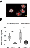
A, Confocal image displaying the distribution of the CB2R mark (red labeling) in the metastatic TNBC line cells, MDA-MB-231, during mitosis and interphase. B, Comparative fluorescence intensity profile of CB2R in mitotic and interphase TNBC cells. Mann-Whitney U test: ****, P < 0.0001. n = 11 and 10 for mitotic HCC-1395 and MDA-MB-231 cells, respectively; whereas n = 37 and 38 for interphase HCC-1395 and MDA-MB-231 cells, respectively.
Fig 4
Fig 4. Treatment of TNBC cells with HU-308 releases CB2R from the nucleoli. (A), confocal micrographs of cancer cell lines after 24 hours in the presence of 10 µM of the selective CB2R agonist, HU-308. Comparative analysis of red channel fluorescence intensity in the nucleolus (B), nucleoplasm (C), and cytoplasm (D). Dunn’s post hoc test: **, P < 0.01 and ****, P < 0.0001. The number of cells evaluated is indicated at the top of the whisker in the box-and-whisker plots of panel B, which are the same for panels C and D.
Fig 5
Fig 5. Effect of the agonist HU-308 on the migration of TNBC cells, assessed with the wound healing assay.
Representative images of the scratch/wound in HCC-1395 (A) and MDA-MB-231 cells (B) at 0 and 12 h. The red lines delimit the edge of the wound at time 0 h. Quantitative analysis of wound closure over the 24 h-period (for simplicity, only the 0-21 h lapse is shown) for HCC-1395 (C) and MDA-MB-231 cells (D). For B and C, unpaired t -test: &, P < 0.05 - 0.01; #, P < 0.05 - 0.0001.
Fig 6
Fig 6. The CB2 receptor agonist HU-308 reduces the proliferation of TNBC metastatic cells.
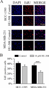
A, Representative images of TNBC cell lines stained with EdU (purple, denoting proliferating cells), DAPI (blue), and the merge of both channels (violet). B, Bar graphs of the cell proliferation, obtained as a percentage from the relative fold of EdU-positive cells. Experiments were conducted in triplicate. For B, unpaired t-test: ***, P < 0.0001.

References

    1. Cuthrell KM, Tzenios N. Breast cancer: updated and deep insights. Int Res J Oncol. 2023;6(1):104–18.
    1. Sung H, Ferlay J, Siegel RL, Laversanne M, Soerjomataram I, Jemal A, et al.. Global cancer statistics 2020: GLOBOCAN estimates of incidence and mortality worldwide for 36 cancers in 185 countries. CA Cancer J Clin. 2021;71(3):209–49. doi: 10.3322/caac.21660 - DOI - PubMed
    1. Bimonte S, Palma G, Cascella M, Cuomo A. Phytocannabinoids in triple negative breast cancer treatment: current knowledge and future insights. Anticancer Res. 2023;43(3):993–1000. doi: 10.21873/anticanres.16243 - DOI - PubMed
    1. Dariš B, Tancer Verboten M, Knez Ž, Ferk P. Cannabinoids in cancer treatment: therapeutic potential and legislation. Bosn J Basic Med Sci. 2019;19(1):14–23. doi: 10.17305/bjbms.2018.3532 - DOI - PMC - PubMed
    1. Dobovišek L, Borštnar S, Debeljak N, Kranjc Brezar S. Cannabinoids and triple-negative breast cancer treatment. Front Immunol. 2024;15:1386548. doi: 10.3389/fimmu.2024.1386548 - DOI - PMC - PubMed

MeSH terms

Substances

LinkOut - more resources