

Development of a Z-score equation for atrioventricular interval measurement by two-dimensional pulsed Doppler echocardiography in normal fetuses between 16 and 33+6 weeks of gestation
- PMID: 40360248
- PMCID: PMC12081140
- DOI: 10.14366/usg.24142
Development of a Z-score equation for atrioventricular interval measurement by two-dimensional pulsed Doppler echocardiography in normal fetuses between 16 and 33+6 weeks of gestation
Abstract
Purpose: Fetal echocardiography is the primary diagnostic tool for assessing the atrioventricular (AV) time interval. Establishing a reference range for this parameter throughout pregnancy is essential for the early detection of potential abnormalities. The aim of this study was to develop a Z-score equation and establish specific percentiles for the AV time interval in normal fetuses between 16 and 33+6 weeks of gestation.
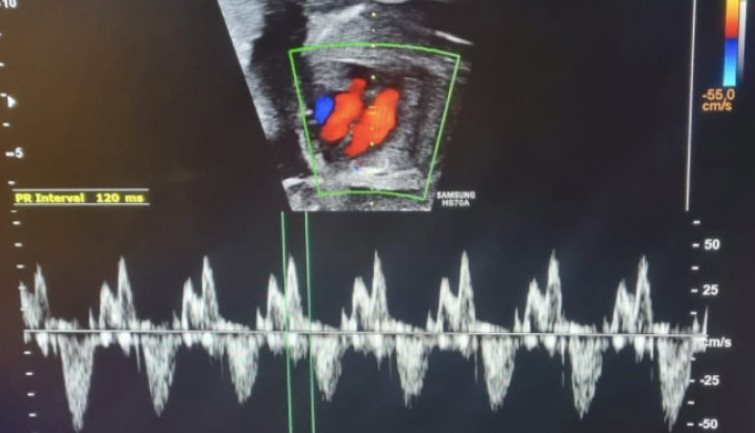
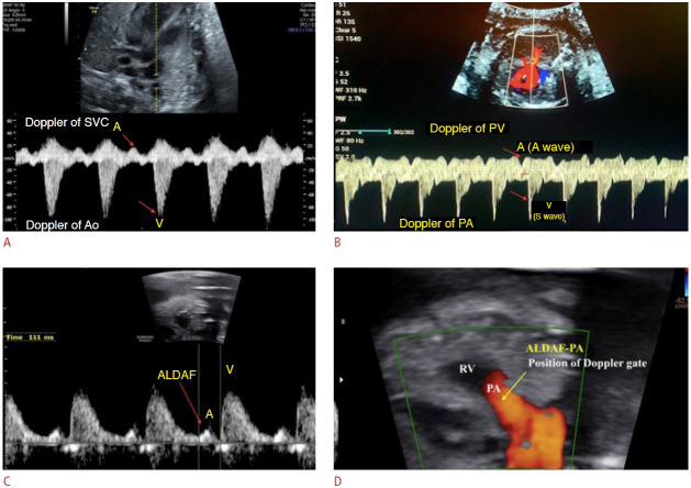
Methods: A multicenter, prospective, cross-sectional study was conducted between 2018 and 2022. A large sample of pregnant women meeting specific eligibility criteria was included, while cases with potential confounders were excluded. Two-dimensional echocardiography with pulsed Doppler techniques was employed, focusing on the left ventricular inflow and outflow. Data were rigorously analyzed with careful assessment of measurements and normalization procedures.
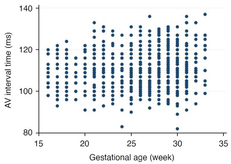
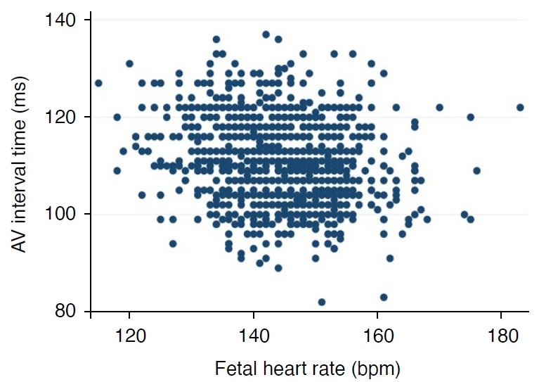
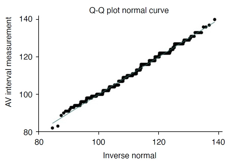
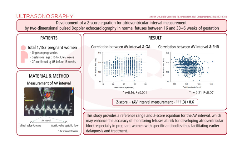
Results: In total, 1,309 echocardiograms were performed, and 1,183 pregnant women were included after applying the eligibility criteria. Detailed percentiles for each gestational age were determined, and a Z-score equation was formulated. A very weak correlation was observed between AV interval measurement and gestational age (r=0.16, P<0.001). In addition, the correlation between AV interval measurement and fetal heart rate was weak (r=-0.21, P<0.001). The Z-score for the AV interval measurement in milliseconds was derived as follows: Z-score=(AV interval measurement-111.3)/8.6.
Conclusion: This study provides a reference range and Z-score equation for the AV interval, which may enhance the accuracy of monitoring fetuses at risk for developing atrioventricular block-especially in pregnant women with specific antibodies-thus facilitating earlier diagnosis and treatment.
Keywords: Atrioventricular interval; Fetal heart; Reference values; Two-dimensional echocardiography.
Conflict of interest statement
No potential conflict of interest relevant to this article was reported.
Figures
References
-
- Donofrio MT, Moon-Grady AJ, Hornberger LK, Copel JA, Sklansky MS, Abuhamad A, et al. Diagnosis and treatment of fetal cardiac disease: a scientific statement from the American Heart Association. Circulation. 2014;129:2183–2242. - PubMed
-
- Hoffman JI, Kaplan S, Liberthson RR. Prevalence of congenital heart disease. Am Heart J. 2004;147:425–439. - PubMed
-
- van der Linde D, Konings EE, Slager MA, Witsenburg M, Helbing WA, Takkenberg JJ, et al. Birth prevalence of congenital heart disease worldwide: a systematic review and meta-analysis. J Am Coll Cardiol. 2011;58:2241–2247. - PubMed
-
- Michaelsson M, Engle MA. Congenital complete heart block: an international study of the natural history. Cardiovasc Clin. 1972;4:85–101. - PubMed
LinkOut - more resources
Full Text Sources
