. 2025 May 13;13(5):e011378.
doi: 10.1136/jitc-2024-011378.

Combination of a therapeutic cancer vaccine targeting the endogenous retroviral envelope protein ERVMER34-1 with immune-oncology agents facilitates expansion of neoepitope-specific T cells and promotes tumor control

Affiliations

Combination of a therapeutic cancer vaccine targeting the endogenous retroviral envelope protein ERVMER34-1 with immune-oncology agents facilitates expansion of neoepitope-specific T cells and promotes tumor control

Maria Del Mar Maldonado et al. J Immunother Cancer. .

Abstract

Background: Endogenous retroviruses (ERVs) are remnants of retrovirus germline infections that occurred over the course of evolution and constitute between 5% and 8% of the human genome. While ERVs tend to be epigenetically silenced in normal adult human tissues, they are often overexpressed in carcinomas and may represent novel immunotherapeutic targets. This study characterizes the ERV envelope protein ERVMER34-1 as a target for a therapeutic cancer vaccine.

Methods: The expression of ERVMER34-1 in multiple healthy adult and cancer tissues was assessed, as was its immunogenicity, to ascertain whether specific T cells could lyse human carcinoma cell lines expressing ERVMER34-1. Furthermore, the ability of a rationally designed ERVMER34-1-targeted therapeutic vaccine to induce tumor clearance in two murine carcinoma models expressing ERVMER34-1 was examined either as a monotherapy or in combination with anti-programmed cell death protein-1/programmed death-ligand 1 monoclonal antibody (mAb) or the interleukin-15 superagonist N-803.

Results: The ERVMER34-1 protein was shown to be overexpressed in 232/376 of human carcinomas analyzed while being absent in most healthy adult tissues. High levels of ERVMER34-1 RNA expression associate with decreased survival in uveal melanoma, adenoid cystic, and head and neck carcinomas. ERVMER34-1-specific T cells were detected in peripheral blood mononuclear cells (PBMCs) of patients with cancer but not healthy donors following an overnight stimulation. However, reactive T cells are readily expanded from both healthy donor and patient with cancer PBMCs following a 7- day in vitro stimulation. Furthermore, ERVMER34-1-specific T cells selectively kill human carcinoma cell lines expressing ERVMER34-1. A novel, rationally designed, therapeutic cancer vaccine targeting ERVMER34-1 mediated tumor control in established syngeneic murine tumors expressing the full-length ERVMER34-1 protein. When combined with checkpoint blockade, the vaccine promoted expansion of neoepitope-reactive T cells whose function was further enhanced when combined with N-803. This expansion of neoepitope-reactive T cells was associated with tumor control.

Conclusions: This study reveals the potential of a vaccine that targets the retroviral envelope protein ERVMER34-1 and supports its continued development toward clinical testing as a new class of therapeutic cancer vaccine.

Keywords: Cytokine; Immune Checkpoint Inhibitor; Immunotherapy; Vaccine.


Conflict of interest statement

Competing interests: The authors have no competing interests to disclose.

Figures
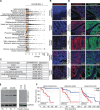
Figure 1
Figure 1
Identification of ERVMER34-1 as a potential therapeutic target. (A) RNA expression levels of ERVMER34-1 in multiple human carcinomas (black dots) along with histologically normal tissues adjacent to the tumor when available (orange dots). The shaded area indicates the median expression of ERVMER34-1 in normal tissues adjacent to the tumor plus 2 SD. The number of samples included in the analysis for each tumor type and corresponding adjacent normal tissues, respectively, were: adrenocortical (79, 0), bladder urothelial (411, 19), breast invasive (1,095, 112), cervical squamous cell and endocervical adenocarcinoma (304, 3), cholangiocarcinoma (35, 9), colon adenocarcinoma (469, 41), esophageal (161, 11), glioblastoma multiforme (155, 5), head and neck squamous cell (500, 45), liver hepatocellular (371, 51), lung adenocarcinoma (525, 59), lung squamous (501, 49), pancreatic (177, 4), pheochromocytoma and paraganglioma (178, 3), prostate (499, 52), rectum adenocarcinoma (166, 10), sarcoma (259, 2), skin cutaneous melanoma (103, 1), stomach adenocarcinoma (375, 32), thyroid (502, 58), thymoma (119, 2), uterine corpus endometrial (547, 35), uveal melanoma (80, 0). (B) Representative images of immunohistochemical (IHC) staining of ERVMER34-1 protein expression (red) in histologically normal human cerebellum, colon, breast, uterus, and testis. Scale bars indicate 100 µm. Blue signal represents DAPI staining. (C) The table shows the number of colon, head and neck, bladder, lung, endometrium, and breast carcinoma tissues demonstrating ERVMER34-1 expression in greater than 10% of tumor cells as assessed by IHC, relative to the total number of tissues evaluated for each tumor type. The percent positive is indicated in parentheses. (D) Representative images of immunohistochemical staining of ERVMER34-1 protein expression (red) in human colon carcinoma, bladder carcinoma, lung adenocarcinoma, lung squamous cell carcinoma, and endometrial carcinoma. Cytokeratin (green) is included as a tumor marker. Scale bars indicate 50 µm. Blue signal represents DAPI staining. (E) Western blot of ERVMER34-1 expression in protein lysates of matched tumor (T) and tumor adjacent (TA) tissues collected from patients diagnosed with either lung (LUAD) n=1 or colon (COAD) adenocarcinomas n=1 (left panel). ERVMER34-1 expression in lung n=1 and colon n=1 protein lysates collected from healthy donors is shown in the right panel. GAPDH is used as a loading control. (F) Survival analysis of patients with cancer was performed using Gene Expression Profiling Interactive Analysis (GEPIA, http://gepia.cancer-pku.cn/) for patients diagnosed with uveal melanoma (n=26 high; n=26 low), head and neck squamous carcinoma (n=170 high; n=169 low) and adenoid cystic carcinoma (n=26 high; n=26 low) based on high (red) and low (blue) ERVMER34-1 RNA expression in tumor tissues. Cut-offs for high and low ERVMER34-1 expression were set to the top 66% and lower 33%, respectively. DAPI, 4’,6-diamidino-2-phenylindole; FPKM, Fragments Per Kilobase per Million mapped fragments; GAPDH, glyceraldehyde-3-phosphate dehydrogenase.
Figure 2
Figure 2
ERVMER34-1-specific immunity can be expanded through in vitro stimulation. (A) Immune reactivity of healthy donor PBMCs (n=13) and patient with cancer PBMCs (patients with breast, n=4; colon, n=4; lung, n=7; and prostate carcinoma, n=4) against the 15-mer ERVMER34-1 peptide library, as assessed using a 16-hour IFN-γ ELISpot assay. *p≤0.05 by an unpaired t-test. (B) Graphical representation of 7-day in vitro expansion of reactive T cells using the ERVMER34-1 peptide library. (C) Representative image of IFN-γ ELISpot plate using PBMCs collected from a patient with breast cancer using the stimulation protocol described in panel B. Each well contains a single 15-mer peptide comprising our ERVMER34-1 peptide library for a total of 93 peptides. Wells outlined in blue are no-peptide DMSO negative control. The well outlined in black is a PMA/ION positive control. Data of responses from an additional 11 donors are presented in online supplemental figure 2B. (D) Mapping the diversity of ERVMER34-1-reactive CD8+ T cells expanded from PBMCs from healthy Donor A using individual 15-mer ERVMER34-1 peptides that comprise our peptide library. (E) Identifying epitopes of ERVMER34-1-reactive CD8+ T cells expanded from healthy Donor A using 9-mer peptides. (F) Mapping the diversity of ERVMER34-1-reactive CD8+ T cells expanded from PBMCs from healthy Donor B using individual 15-mer ERVMER34-1 peptides that comprise our peptide library. (G) Identifying epitopes of ERVMER34-1-reactive CD8+ T cells expanded from healthy Donor B using 9-mer peptides. (H) Immuno-fluorescent analysis of ERVMER34-1 expression (green signal) in either parental SW620 or SW620 ERVMER34-1 CRISPR knock-out cell line. Blue signal represents DAPI staining. Scale bars indicate 5 µm. (I) Percent specific lysis of the parental SW620 and SW620 ERVMER34-1 CRISPR knockout cell lines following overnight incubation with ERVMER34-1-specific CD8+ T cells expanded from PBMCs collected from healthy Donor C and using an effector-to-target ratio of 5:1. ****p≤0.0001 by an unpaired t-test. (J) Western blot of ERVMER34-1 protein expression in multiple human carcinoma cell lines; GAPDH used as a protein loading control. (K) Lysis of indicated human carcinoma cell lines over time using ERVMER34-1-specific CD8+ T cells expanded from healthy Donor B using an effector-to-target ratio of 10:1. DAPI, 4’,6-diamidino-2-phenylindole; DMSO, dimethyl sulfoxide; ELISpot, Enzyme-Linked ImmunoSpot; GAPDH, glyceraldehyde-3-phosphate dehydrogenase; IFN, interferon; PBMCs, peripheral blood mononuclear cells; PMA/ION, phorbol 12-myristate 13-acetate/ionomycin.
Figure 3
Figure 3
Generation of a therapeutic cancer vaccine targeting ERVMER34-1. (A) Amino acid sequence of ERVMER34-1 with identified protein regions highlighted. The sequences removed from the ERVMER34-1 vaccine are underlined. (B) Enumeration of ERVMER34-1-reactive T cells in splenocytes harvested from non-tumor-bearing BALB/c and C57BL/6 mice vaccinated on days 0 and 7 with either control adenovirus or ERVMER34-1 vaccine using a 16-hour IFN-γ ELISpot assay performed 2 weeks following the last vaccine dose. Each row is an individual animal, and each column is a single peptide comprising the 15-mer peptide library (n=3 mice per group). (C) Enumeration of ERVMER34-1-specific multifunctional T cells in non-tumor-bearing C57BL/6 mice following vaccination with either control adenovirus or the ERVMER34-1 vaccine as assessed by an ex vivo intracellular cytokine production assay. Number indicates the percentage of total TCRbeta+ CD8+ T cells secreting both TNF-α and IFN-γ in response to ERVMER34-1 peptides (n=10 mice per group). Mice were treated as described above. Data representative of two independent experiments. (D) Immunofluorescence staining of ERVMER34-1 protein expression (green signal) in MC38 parental and MC38 pERVMER34-1 tumors. Blue signal represents DAPI staining; scale bars indicate 100 µm. (E) Tumor growth curves of MC38 pERVMER34-1 tumors in mice that received either no treatment (NT) or a single dose (1010 viral particles, subcutaneously) of either control adenovirus or ERVMER34-1 vaccine. Mice were randomized and treated when tumors reached a size of 50–100 mm3 (n=9–10 mice per group). Data representative of two independent experiments. (F) Survival curves of mice bearing MC38 pERVMER34-1 tumors that received no treatment or one dose of either control adenovirus or ERVMER34-1 vaccine. (G) Flow cytometry analysis of tumor-infiltrating lymphocytes in mice bearing MC38 ERVMER34-1 tumors treated with control adenovirus or ERVMER34-1 vaccine (n=5 mice per group). Immune readout was performed 6 days post-vaccination. (H) Enumeration of specific ERVMER34-1-reactive cells present in expanded tumor- infiltrating lymphocytes as assessed by an IFN-γ ELISpot assay using the ERVMER34-1 peptide library as a source of antigen. (I) ERVMER34-1 immune reactivity of splenocytes and expanded tumor-infiltrating lymphocytes harvested from mice vaccinated with control adenovirus or ERVMER34-1 vaccine as assessed by an IFN-γ ELISpot assay using individual 15-mer ERVMER34-1 peptides that comprise our library (n=5 mice per group). (J) Comparison of immune reactivity of MC38 neoepitopes, and p15e of tumor-infiltrating T cells in mice bearing MC38 pERVMER34-1 tumors treated with either control adenovirus or ERVMER34-1 vaccine (n=4–5 mice per group). (K) Assessment of IFN-γ present within the tumor microenvironment in mice treated with ERVMER34-1 vaccine (n=3–4 mice per group). (L) Expression of PD-L1 on the surface of CD45 cells (left panel) and CD45+CD11b+ cells (right panel) within the MC38 pERVMER34-1 tumors of mice treated with either control adenovirus or ERVMER34-1 vaccine. *p≤0.05; **p≤0.01; ***p≤0.001 by an unpaired t-test. DAPI, 4’,6-diamidino-2-phenylindole; ELISpot, Enzyme-Linked ImmunoSpot; IFN, interferon; PD-L1, programmed death-ligand 1; TCR, T-cell receptor; TIL, tumor-infiltrating T cell; TNF, tumor necrosis factor.
Figure 4
Figure 4
Combination of immune checkpoint blockade with ERVMER34-1 vaccine enhances its antitumor efficacy. (A) Graphical representation of tumor treatment schedule. (B) Tumor growth curves of MC38 pERVMER34-1 tumors treated with either control adenovirus or ERVMER34-1 vaccine with or without the addition of anti-PD-L1 as indicated in panel A (n=9–10 mice per group). Data representative of two independent studies. (C) Survival curves of mice treated as indicated in panel A. (D) Immune reactivity of splenocytes harvested from mice treated with either control adenovirus or ERVMER34-1 vaccine with or without the addition of anti-PD-L1 as indicated in panel A using a 16-hour IFN-γ ELISpot assay with individual 15-mer ERVMER34-1 peptides in the assay (n=5 mice per group). (E) Immune reactivity of splenocytes harvested from mice treated with either control adenovirus or ERVMER34-1 vaccine with or without the addition of anti-PD-L1 as indicated in panel A using a 16-hour IFN-γ ELISpot assay with individual MC38 neoepitope peptides in the assay (n=5 mice per group). (F) Volcano plot of changes in cytokines and chemokines present within the tumor microenvironment in mice treated as indicated in panel A as assessed by the Olink Target 48 Cytokine assay (n=5 mice per group). (G) Graphical representation of tumor treatment schedule. (H) Growth curves of contralateral MC38 and MC38 pERVMER34-1 tumors in mice treated as indicated in panel G. Green line: control adenovirus, red line: control adenovirus+anti-PD-1, black line: ERVMER34-1 vaccine, and blue line: ERVMER34-1 vaccine+anti-PD-1 (n=9 mice per group). Data representative of two independent experiments. *p≤0.05; **p≤0.01; ***p≤0.001; ****p≤0.0001 by a two-way analysis of variance followed by a Tukey’s multiple comparisons test when comparing ≥3 groups. ELISpot, Enzyme-Linked ImmunoSpot; IFN, interferon; PBS, phosphate-buffered saline; PD-1, programmed cell death protein-1; PD-L1, programmed death-ligand 1.
Figure 5
Figure 5
Vaccine targeting ERVMER34-1 in combination with immune-oncology agents enhances tumor control. (A) Immunofluorescence staining of ERVMER34-1 protein expression (green signal) in EMT6 parental and EMT6 pERVMER34-1 tumors. Blue signal represents DAPI staining. Scale bars indicate 100 µm. (B) Treatment scheme for panels C–I with mice being treated with either control adenovirus or the ERVMER34-1 vaccine. (C) Tumor growth curves of mice treated as indicated in panel B (n=16–18 mice per group). Data representative of two independent studies. *p≤0.05; **p≤0.01; ****p≤0.0001 by a two-way ANOVA followed by a Tukey’s multiple comparisons test. (D) ERVMER34-1 immune reactivity of splenocytes harvested from mice vaccinated with control adenovirus or ERVMER34-1 vaccine as assessed by a 16-hour IFN-γ ELISpot assay (n=10 mice per group). ****p≤0.0001 by an unpaired t-test. (E) Enumeration of multifunctional T cells in splenocytes specific for a pool of neoepitopes expressed in the EMT6 tumor model (right panel). Control non-mutated neoepitope immune responses are presented in the left panel. (F) Flow cytometry assessment of CD8 effector cells in the tumor of mice treated as indicated. Low and high neoepitope immunity mice are segregated as indicated in panel E by an unpaired t-test. (G) Volcano plot of differences in cytokines and chemokines present within the TME assessed in high versus low neoepitope immune responses by the Olink Target 48 cytokine assay. (H) Tumor growth curves of mice treated with ERVMER34-1 vaccine+anti-PD-L1, which had been identified in panel E as having either a high or low neoepitope immune response as compared with mice treated with control adenovirus+anti-PD-L1 using a two-way ANOVA followed by a Tukey’s multiple comparisons, *p≤0.05; **p≤0.01. (I) Correlation analysis of neoepitope-reactive T cells in the EMT6 pERVMER34-1 tumor model with tumor volume of mice treated as indicated in panel B. (J) Treatment scheme for panels K–M with mice being treated with either control adenovirus or the ERVMER34-1 vaccine. (K) Spider plots of changes in tumor volume of EMT6 pERVMER34-1 tumors treated as indicated in panel J (n=10–14 mice per group). Shaded region indicates greater than 20% decrease in tumor size. Data representative of two independent studies. (L) Enumeration of ERVMER34-1-reactive T cells in splenocytes from mice treated as indicated in panel J as assessed by a 16-hour IFN-γ ELISpot assay using the ERVMER34-1 peptide library. Mice that received the triplet regimen that were unresponsive to treatments are labeled as non-responders (NR), whereas those that showed tumor shrinkage greater than 20% are labeled as responders (R). (M) Enumeration of neoepitope reactive T cells in splenocytes from mice treated as indicated in panel J as assessed by intracellular cytokine staining. Mice that received the triplet regimen that were unresponsive to treatments are labeled as NR, whereas those that showed tumor shrinkage greater than 20% are labeled as R. *p≤0.05; **p≤0.01; ***p≤0.001; ****p≤0.0001 by an unpaired t-test by comparing two groups. ANOVA, analysis of variance; ELISpot, Enzyme-Linked ImmunoSpot; IFN, interferon; PBS, phosphate-buffered saline; PD-1, programmed cell death protein-1; PD-L1, programmed death-ligand 1; TNF, tumor necrosis factor.

Similar articles

Cited by

  • The role of HERV envelope protein in ovarian cancer.
    Zhang J, Sun D, Zhan Y, Gao Q, Bao C, Xiang H, Shen Y, Gao Q, Zhang M, Wang J, Mao L. Zhang J, et al. Front Cell Dev Biol. 2025 Jul 31;13:1618542. doi: 10.3389/fcell.2025.1618542. eCollection 2025. Front Cell Dev Biol. 2025. PMID: 40823530 Free PMC article. Review.

References

    1. Feschotte C, Gilbert C. Endogenous viruses: insights into viral evolution and impact on host biology. Nat Rev Genet 2012;13:283–96. 10.1038/nrg3199 - DOI - PubMed
    1. Johnson WE. Origins and evolutionary consequences of ancient endogenous retroviruses. Nat Rev Microbiol 2019;17:355–70. 10.1038/s41579-019-0189-2 - DOI - PubMed
    1. Lander ES, Linton LM, Birren B, et al. Initial sequencing and analysis of the human genome. Nature New Biol 2001;409:860–921. 10.1038/35057062 - DOI - PubMed
    1. Jern P, Coffin JM. Effects of retroviruses on host genome function. Annu Rev Genet 2008;42:709–32. 10.1146/annurev.genet.42.110807.091501 - DOI - PubMed
    1. de Parseval N, Heidmann T. Human endogenous retroviruses: from infectious elements to human genes. Cytogenet Genome Res 2005;110:318–32. 10.1159/000084964 - DOI - PubMed

MeSH terms

Substances