. 2025 May 13;11(1):234.
doi: 10.1038/s41420-025-02405-z.

O-GlcNAcylation of NONO regulates paraspeckle component assembly and contributes to colon cancer cell proliferation

Affiliations

O-GlcNAcylation of NONO regulates paraspeckle component assembly and contributes to colon cancer cell proliferation

Yeolhoe Kim et al. Cell Death Discov. .

Abstract

Non-POU domain-containing octamer-binding protein (NONO) is a multifunctional member of the Drosophila behavior/human splicing (DBHS) protein family with DNA- and RNA-binding activity. NONO is highly expressed in various types of cancer, and excessive O-GlcNAcylation has also been implicated in tumorigenesis. Although recent studies revealed that NONO is O-GlcNAcylated and that this modification is involved in DNA damage repair, it remains unknown whether O-GlcNAcylation of NONO regulates cancer cell proliferation. Additionally, little is known about the effect of O-GlcNAcylation on other biological properties of NONO. In this study, we identify Thr440 as the primary NONO O-GlcNAcylation site and demonstrates its crucial role in the assembly of paraspeckles, an important subnuclear compartment that facilitates NONO-dependent transcriptional regulation in mammalian cells. Moreover, we found that O-GlcNAcylation of NONO is required to maintain the expression of genes related to microtubule cytoskeleton organization involved in mitosis and to suppress the expression of genes related to cellular response to type I interferon. Regarding the regulation of these genes, depletion of NONO O-GlcNAcylation at Thr440 significantly inhibited the proliferation of colon cancer cells. Collectively, our findings highlight NONO O-GlcNAcylation as a key regulator modulating paraspeckle formation and as a candidate therapeutic target in colon cancer.


Conflict of interest statement

Competing interests: The authors declare no competing interests. Ethics approval: All methods were performed in accordance with the relevant guidelines and regulations. All experimental protocols were approved by the The Institutional Animal Care and Use Committees of the Laboratory Animal Research Center at Yonsei University (Approval Numbers: IACUC-A-202107-1286-02, IACUC-A-202205-1470-01). Our manuscript does not contain any human data.

Figures
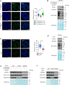
Fig. 1
Fig. 1. The number of NONO-containing paraspeckle-like structures decreases by lowering cellular O-GlcNAcylation.
A NONO immunostaining on colon cancer cell HCT116 in 25 mM glucose, 5.5 mM glucose, or 5.5 mM glucose transfected with myc-tagged OGT conditions. B Cellular O-GlcNAcylation status was detected via western blot. C NONO immunostaining on colon cancer cell HCT116 in siControl, or siOGT conditions. D Cellular O-GlcNAcylation status was detected via western blot. E, F Nuclear and cytoplasmic fractions were analyzed by western blot with relevant antibodies. Lamin A/C and HSP90α/β were used as nuclear and cytoplasmic markers, respectively. Results in (A), (C) are represented as boxes and whiskers: 10-90 percentile range, “+” sign represents mean, n > 50 nuclei from 3 different biologically independent replicates. ***P < 0.001 (A; one-way ANOVA with Tukey’s multiple comparisons test, C; Unpaired two-tailed t-test). Scale bar represents 5 μm.
Fig. 2
Fig. 2. NONO O-GlcNAcylation site was identified as Thr440.
A sWGA precipitation was performed to evaluate immunoprecipitated NONO level upon with or without GlcNAc competition. Immunoprecipitated NONO protein level was tested by western blot analysis. B Validation of O-GlcNAcylation with endogenously-expressed NONO in HCT116 cells. The NONO O-GlcNAcylation in HCT116 was higher in cells transfected with OGT. C The NONO O-GlcNAcylation in HCT116 was lower in cells transfected with OGA. D A co-IP assay was performed to evaluate the interactions between NONO with OGT. HCT116 WCLs were subjected to IP with normal rabbit IgG of anti-NONO antibody. E Mass spectrometry (MS) analysis identifies the O-GlcNAcylation site in NONO. EThcD spectra of O-Glycopeptide FGQAATMEGIGAIGGTPPAFNR from human NONO is shown. The site of O-GlcNAc modification was identified as threonine 440. The c and z fragments detected are as indicated in the sequence. F Human NONO protein structure. O-GlcNAcylation was indicated at threonine 440. (RRM; RNA recognition motif, NOPS; NONA/Paraspeckle). G IP assay to compare the O-GlcNAcylation levels of wild-type NONO with T440 to A mutant. 10 ug of either FLAG-tagged NONO WT or mutant were transfected into HEK293 cells and lysed after 24 h. NONO O-GlcNAcylation level was normalized to the immunoprecipitated NONO protein level (n = 3 per condition). H Sequence information indicates that NONO threonine 440 is a highly conserved sequence in mammals. Data are presented as mean ± SD; ***P < 0.001, Unpaired two-tailed t-test.
Fig. 3
Fig. 3. Thr440 O-GlcNAcylation of NONO is necessary for paraspeckle components interaction.
A, B A co-IP assay was performed to evaluate the interactions between NONO with SFPQ, and PSPC1. HCT116 cells were treated with OSMI4 or siOGT and lysed after 24 h or 48 h, respectively. WCLs were subjected to IP with anti-NONO antibody. Relative co-immunoprecipitated SFPQ, PSPC1 level was normalized to NONO (n = 3 per condition). C NONO immunostaining on NONO knockout, stably recovered with NONO WT, or T440A colon cancer cell HCT116. n > 50 nuclei from 3 different biologically independent replicates. D O-GlcNAcylation levels of stably expressed WT NONO with T440A mutant were compared (n = 3 per condition). E, F A co-IP assay was performed to evaluate the interactions between NONO WT or T440A with SFPQ, and PSPC1. NONO KO HCT116 cells were transfected with FLAG-tagged NONO WT or T440A and transfected with myc-tagged SFPQ or PSPC1 and lysed after 24 h. Immunoprecipitation was performed with FLAG®-agarose. Relative co-immunoprecipitated SFPQ, PSPC1 level was normalized to NONO WT or T440A (n = 3 per condition). G RIP analysis was performed to evaluate co-immunoprecipitated lncRNA NEAT1_2 level with NONO WT or T440A. Schematic of RNA immunoprecipitation experimental procedure. H Total NEAT1_2 level was evaluated by qPCR analysis (n = 3 per condition). I Co-immunoprecipitated NEAT1_2 level with NONO WT or T440A protein was evaluated by qPCR analysis (n = 3 per condition). Data are presented as mean ± SD; **P < 0.01, ***P < 0.001, Unpaired two-tailed t-test. Scale bar represents 5 μm.
Fig. 4
Fig. 4. Modifying NONO O-GlcNAcylation status results in transcriptional profile alteration.
A, B GSEA(Gene Set Enrichment Analysis) plots of biological functions associated with gene groups whose expression changes in NONO knockdown by siRNA. C Volcano plot for differentially expressed gene (DEG) analysis. Genes related to microtubule cytoskeleton organization were downregulated in NONO knock-downed HCT116. Conversely, genes related to cellular response to type I interferon were upregulated in same condition. D, E qPCR was performed to evaluate the genes associated with microtubule cytoskeleton organization involved in mitosis or cellular response to Type I interferon (n = 3 per condition). Data are presented as mean ± SD; *P < 0.05, **P < 0.01, ***P < 0.001 ((D, E); one-way ANOVA with Tukey’s multiple comparisons test).
Fig. 5
Fig. 5. Thr440 O-GlcNAcylation of NONO promotes cancer cell invasion, colony formation, and proliferation.
A WST-8 assay was performed to compare cell proliferation between NONO knockout, NONO WT-expressing, or NONO T440A-expressing HCT116 cells (n = 3 per condition). B Transwell invasion assay was performed and invaded cells were measured by WST-8 assay (n = 3 per condition). C NONO knockout, NONO WT-expressing, or NONO T440A-expressing HCT116 cells were cultured for soft agar colony formation assay. Colony formation was examined under a light microscope. Colony formation was measured by using a 485/520 nm filter set (n = 5 per condition). D BALB/c nude mice (n = 6 per group) were injected with 1 × 107 of the indicated cells subcutaneously. Tumor volume and weight were measured 60 days after injection. Data are presented as mean ± SD; *P < 0.05, **P < 0.01, ***P < 0.001, one-way ANOVA with Tukey’s multiple comparisons test.
Fig. 6
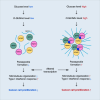
Fig. 6. Schematic of the proposed model depicting O-GlcNAcylation of NONO at Thr440 and its role in cancer cell proliferation.
In cancer cells, a relatively high level of glucose allows more cellular O-GlcNAc modification. O-GlcNAcylation of NONO at Thr440 enhances the paraspeckle formation. Upregulated paraspeckle formation promotes transcriptional alteration, leading to cancer cell proliferation.

Similar articles

References

    1. Passon DM, Lee M, Rackham O, Stanley WA, Sadowska A, Filipovska A, et al. Structure of the heterodimer of human NONO and paraspeckle protein component 1 and analysis of its role in subnuclear body formation. Proc Natl Acad Sci USA. 2012;109:4846–50. 10.1073/pnas.1120792109. - PMC - PubMed
    1. Knott GJ, Bond CS, Fox AH. The DBHS proteins SFPQ, NONO and PSPC1: a multipurpose molecular scaffold. Nucleic Acids Res. 2016;44:3989–4004. 10.1093/nar/gkw271. - PMC - PubMed
    1. Fox AH, Lam YW, Leung AKL, Lyon CE, Andersen J, Mann M, et al. Paraspeckles: a novel nuclear domain. Curr Biol. 2002;12:13–25. 10.1016/S0960-9822(01)00632-7. - PubMed
    1. Fox AH, Lamond AI. Paraspeckles. Cold Spring Harb Perspect Biol. 2010;2:687–8. 10.1101/cshperspect.a000687. - PMC - PubMed
    1. Fox AH, Nakagawa S, Hirose T, Bond CS. Paraspeckles: where long noncoding RNA meets phase separation. Trends Biochemical Sci. 2018;43:124–35. 10.1016/j.tibs.2017.12.001. - PubMed

LinkOut - more resources