. 2025 May 14;10(1):157.
doi: 10.1038/s41392-025-02245-4.

Exosomal transfer of pro-pyroptotic miR-216a-5p exacerbates anthracycline cardiotoxicity through breast cancer-heart pathological crosstalk

Affiliations

Exosomal transfer of pro-pyroptotic miR-216a-5p exacerbates anthracycline cardiotoxicity through breast cancer-heart pathological crosstalk

Yan Ma et al. Signal Transduct Target Ther. .

Abstract

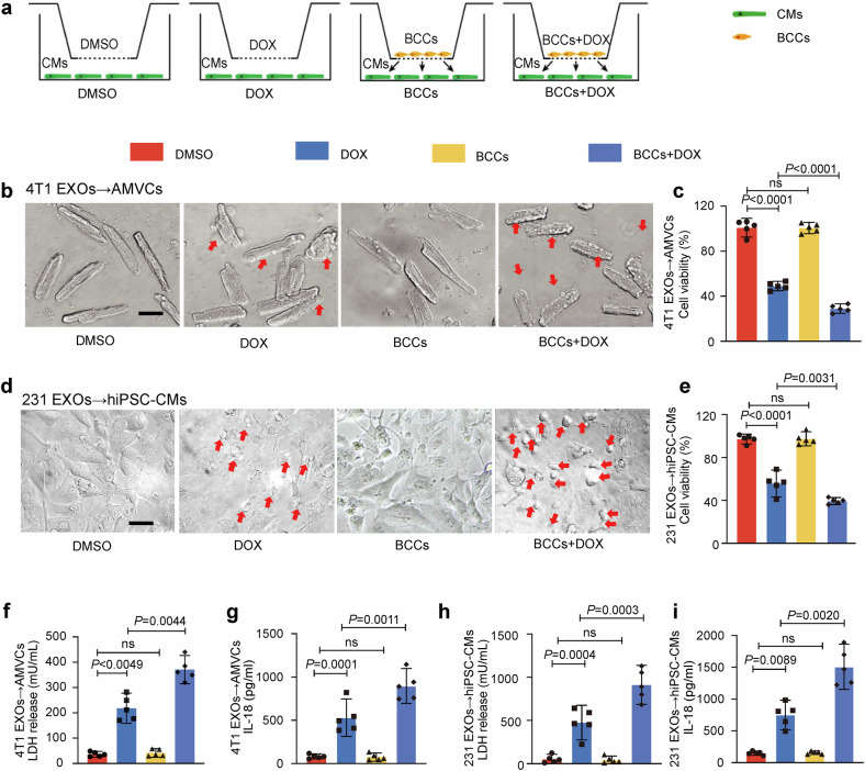
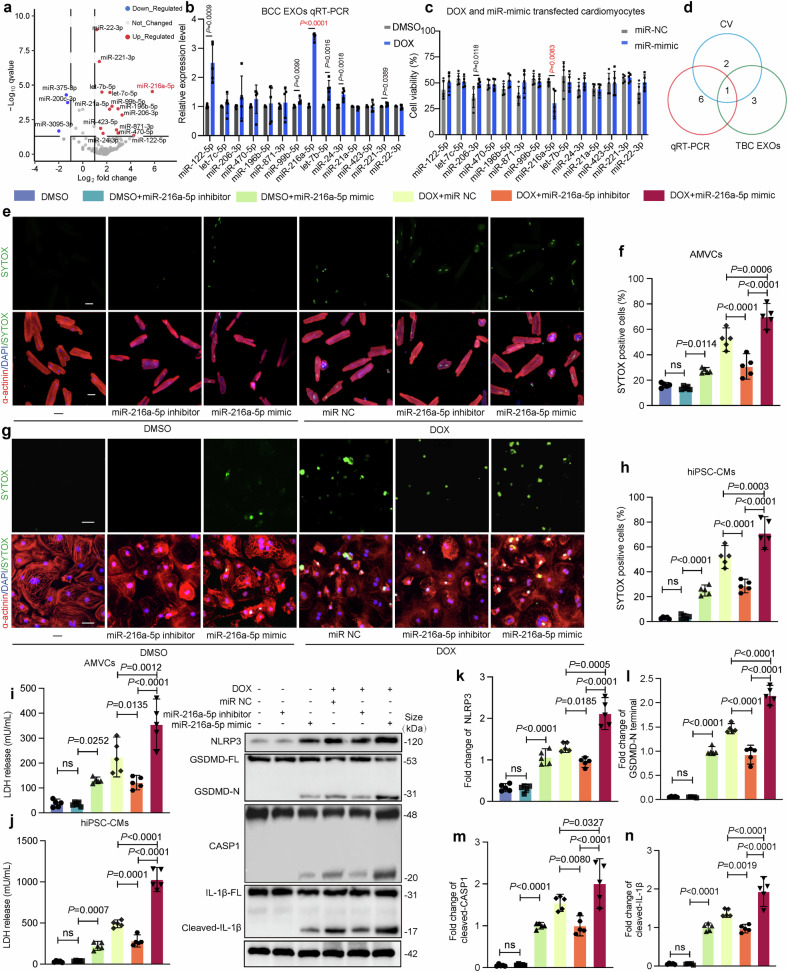
Doxorubicin (DOX) is the most effective chemotherapeutic for breast cancer, but it is usually associated with severe cardiotoxicity. Further investigation to alleviate its side effects is essential. The present study investigated the mechanism of the cross-organ communication between tumors and the heart and potential intervention targets. Morphological bubble-like protrusions were observed in both adult murine ventricular cardiomyocytes (AMVCs) and human induced pluripotent stem cell-derived cardiomyocytes (hiPSC-CMs) cocultured with breast cancer cells (BCCs), along with elevated expression of pyroptosis-related proteins. Exosomes (EXOs) from DOX-treated BCCs aggravated DOX-induced cardiotoxicity (DOXIC) in an orthotopic mouse model of breast cancer. Blocking miRNAs by knocking down Rab27a or inhibiting the release of EXOs in cancer tissue by Dicer enzyme knockout attenuated this additional injury effect. Exosomal miRNA sequencing revealed that miR-216a-5p is especially upregulated in EXOs from DOX-induced BCCs. Mechanistically, miR-216a-5p was upregulated by enhanced transcription mediated by DOX-induced AMP-dependent transcription factor 3 (ATF3) and packaged into EXOs by splicing factor 3b subunit 4 (SF3B4) in BCCs. Itchy E3 ubiquitin-protein ligase (ITCH) was identified as a novel downstream target mRNA of miR-216a-5p. ITCH negatively mediated thioredoxin-interacting protein (TXNIP) ubiquitination to activate the NOD-, LRR- and pyrin domain-containing protein 3 (NLRP3) inflammasome pathway, ultimately leading to cardiomyocyte pyroptosis. Our findings revealed novel cross-organ pathogenic communication between breast cancer and the heart through the exosomal miR-216a-5p-mediated ITCH/TXNIP/NLRP3 pathway, which drives cardiomyocyte pyroptosis. These findings suggest that targeting myocardial miR-216a-5p or blocking harmful EXOs from breast cancer is a potential therapeutic strategy for alleviating DOXIC.


Conflict of interest statement

Competing interests: The authors declare no competing interests.

Figures

Fig. 1
Fig. 1
Coincubation with breast cancer cells aggravates DOX-induced pyroptosis in cardiomyocytes. Adult murine ventricular cardiomyocytes (AMVCs) and human induced pluripotent stem cell-derived cardiomyocytes (hiPSC-CMs) were cocultured with breast cancer cells (BCCs) for 24 hours with 1 μM DOX treatment. a Schematic diagram of the coculture of BCCs with cardiomyocytes using transwell chambers. be Representative images of and statistics for cell viability after DOX treatment. Scale bar, 50 μm.; (f, g) lactate dehydrogenase (LDH) release and IL-18 levels were assessed using colorimetric methods in AMVCs (n = 5) and in (h and i) hiPSC-CMs (n = 5) (one-way ANOVA was used to compare all experimental groups to the DMSO group). “BCCs” indicates breast cancer cells, Directional arrows indicate exosome transfer between donor cells (4T1 or MDA-MB-231) and recipient cells (hiPSC-CMs or AMVCs). “N-BCC-EXOs” denotes normal breast cancer cell EXOs, and “D-BCC-EXOs” denotes DOX-induced breast cancer cell EXOs. “ns” indicates non-significant, and “DOX” indicates doxorubicin. Data are presented as Mean ± SD
Fig. 2
Fig. 2
Exosomes from breast cancer cells exacerbate DOX-induced cardiomyocyte pyroptosis in vitro. a EXOs from BCCs were characterized using transmission electron microscopy (TEM). Scale bar: 100 nm. b Particle size distribution was determined using NanoSight tracking analysis. (NTA). EXO particle concentration in the cell culture supernatant with or without DOX. c Western blot analysis of HSP70, CD81, TSG101, GM130, and Calnexin in cell lysate and 4T1-derived EXOs with or without DOX. d In vitro and ex vivo breast cancer EXO uptake analysis showing fluorescence-labeled EXOs internalized by human induced pluripotent stem cell-derived cardiomyocytes (hiPSC-CMs) or adult murine ventricular cardiomyocytes (AMVCs) observed under a confocal microscope. For in vitro analysis, PKH67-labeled EXOs were harvested and subsequently incubated with hiPSC-CMs. For ex vivo analysis, AMVCs were isolated from mouse hearts 24 hours after tail vein injection of PKH67-labeled EXOs. Scale bar: 20 μm. e, f Representative images of AMVCs and hiPSC-CMs after DOX + N-BCC-EXO or DOX + D-BCC-EXO treatment for 24 hours and (g, h) assessments of cell viability, (i, j) CASP1 expression levels, and (k, l) LDH release levels (n = 5) are shown. Top row: bright-field images, scale bar: 50μm; middle row: CASP1 immunofluorescence (green), scale bar: 20 μm; bottom row: merged images of α-actinin (red), CASP1 (green), and DAPI (blue), scale bar: 20 μm. m Pyroptosis-related protein levels in AMVCs were assessed via Western blotting. All the experimental groups were compared with the DOX group via one-way ANOVA. n IL-18 levels in AMVCs were determined using a colorimetric method (n = 5). “N-BCC-EXOs” denotes normal breast cancer cell EXOs, and “D-BCC-EXOs” denotes DOX-induced breast cancer cell EXOs. Red represents α-actinin, green represents CASP1, and blue represents DAPI. “DOX” indicates doxorubicin, and “ns” indicates non-significant. Data are presented as Mean ± SD
Fig. 3
Fig. 3
Exosomes from breast cancer cells exacerbate DOX-induced cardiomyocyte pyroptosis in vivo. a Schematic illustration of the subchronic DOX exposure model in mice to test the injury efficacy of D-BCC-EXOs (n = 5). b Representative images of M-mode echocardiography and blood flow velocity (obtained using a Doppler system) in mice. c, d The E/A and the E/E’ ratios were quantified using the Doppler echocardiography. e, f Quantification of the left ventricular (LV) ejection fraction and LV fractional shortening via M-mode echocardiography (n = 5). g Representative images of hematoxylin-eosin (H&E) staining (upper), Sirius red staining indicating myocardial fibrosis (middle), and wheat germ agglutinin (WGA) immunofluorescence staining (lower). Scale bar: 50 μm. h Statistics of vacuolization in ventricular tissues. i The fibrotic area per left ventricle was quantified via Sirius red staining (n = 5). j Cell size was quantified via WGA immunofluorescence staining (n = 5). k Mouse plasma brain natriuretic peptide (BNP) was measured using a colorimetric method (n = 5). lo Pyroptosis-related protein levels in mouse ventricular tissue were assessed via western blotting (n = 5) (one-way ANOVA was used to compare all experimental groups to the DOX group). “E” indicates early diastolic transmitral flow velocity, “E′” indicates early diastolic mitral annular velocity, and “A” indicates late (atrial) diastolic transmitral flow velocity. D-BCC-EXOs, denotes doxorubicin-treated breast cancer EXOs, “N-BCC-EXOs” denotes normal breast cancer cell EXOs, “D-BCC-EXOs” denotes DOX-induced breast cancer cell EXOs, “ns” indicates non-significant, and “DOX” indicates doxorubicin. Data are presented as means ± SD
Fig. 4
Fig. 4
Exosomal miR-216a-5p upregulation induced by DOX aggravates DOX-mediated cardiomyocyte pyroptosis. a Sequencing data of mouse plasma EXO miRNAs were used to identify miRNAs that were differentially expressed. The differentially expressed miRNAs are depicted in a volcano plot. b 4T1 cells were treated with DOX or DMSO, and upregulated exosomal miRNAs were identified by qRT‒PCR (n = 5). c Adult murine ventricular cardiomyocytes (AMVCs) were transfected with a miR-216a-5p mimic/scramble before being treated with DOX. Cell viability was determined by CCK-8 assays (n = 5). d Venn analysis revealed that miR-216a-5p satisfied two conditions. eh miR-216a-5p knockdown and overexpression in cardiomyocytes were achieved through transfection with miR-216a-5p inhibitors and mimics, respectively. Multiple immunofluorescence stains and statistics were used to quantify AMVC and human induced pluripotent stem cell-derived cardiomyocytes (hiPSC-CM) death. DAPI (blue), SYTOX (green), and α-actinin (red) (n = 5). Scale bar: 50 μm. i, j LDH release levels and cell viability were measured (n = 5). kn The protein expression levels of NLRP3, GSDMD-N, cleaved CASP1, and cleaved IL-1β were detected via Western blotting (n = 5) (one-way ANOVA was used to compare all experimental groups with the DOX+miR NC group). “BCCs” indicates breast cancer cells, “ns” indicates non-significant, and “DOX” indicates doxorubicin. Data are presented as means ± SD
Fig. 5
Fig. 5
DOX upregulates ATF3 to promote miR-216a-5p transcription in BCCs. a Relative pri-miR-216a expression in mouse cancer tissue and 4T1 cells from DMSO- and DOX-induced mice was analyzed via qRT-PCR. b Protein levels were assessed by Western blotting using DOX/DMSO-treated 4T1 cells with or without siATF3. c The relative pri-miR-216a expression in DOX/DMSO-treated 4T1 cells with ATF3 overexpression or knockdown was analyzed via qRT-PCR. d, e The direct effects of miR-216a-5p on mouse ATF3 were identified using a luciferase assay. 4T1 cells were cotransfected with siATF3/oeATF3 and reporter plasmids for 48 hours. Firefly/Renilla luciferase activity was used to evaluate regulatory effects. In each group, the fold change was calculated by dividing the firefly/Renilla luciferase activity by the value obtained for the DMSO group. f ChIP-PCR was performed using an anti-ATF3 antibody or IgG in 4T1 cells, and PCR of the miR-216a promoter was performed. The values correspond to the ratio of the anti-ATF3 immunoprecipitated DNA relative to the IgG immunoprecipitated DNA. g ATF3 and miR-216a-5p expression levels gradually increased in a time-dependent manner after DOX treatment, and the two indicators were positively correlated. h Adult murine ventricular cardiomyocytes (AMVCs) cell viability was determined using CCK-8 assays (n = 5). ik LDH release and IL-18 and IL-1β levels in AMVCs were assessed using a colorimetric method (n = 5). “siNC-BCC-EXOs” denotes exosomes from control siRNA-transfected DOX-induced 4T1 cells, “siATF3-BCC-EXOs” denotes exosomes from ATF3-knockdown DOX-induced 4T1 cells, and “DOX” indicates doxorubicin. Data are presented as means ± SD
Fig. 6
Fig. 6
SF3B4 (sap-49) packages miR-216a-5p into 4T1 exosomes. a Three candidate proteins were screened from RBPBD. b The effect of SF3B4 knockdown in 4T1 cells was assessed via western blotting. c The miR-216a-5p level in cell lysates and EXOs was analyzed via qRT‒PCR. d The SF3B4 binding motif and the matched miR-216a-5p sequence. e The binding of the wild-type and mutated miR-216a-5p probes to SF3B4 in 4T1 cells and EXOs from 4T1 cells was measured using an RNA pull-down assay. f The identified motif was the genuine binding region of miR-216a-5p and SF3B4, and mutated versions of miR-216a-5p were tested for binding to SF3B4 via EMSA. g SF3B4 in 4T1 cells was knocked down, and EXOs were isolated. After adult murine ventricular cardiomyocytes (AMVCs) were incubated with 4T1-derived EXOs containing Cy3-miR-216a-5p, Cy3 fluorescence in AMVCs was detected. Scale bar: 20 μm. h AMVCs were treated with SF3B4-knockdown and DOX-exposed 4T1 BCC-EXOs and exposed to DOX. The viability of AMVCs was assessed using CCK-8 assays (n = 5). ik LDH release and IL-18 and IL-1β levels in AMVCs were assessed using a colorimetric method (n = 5). “siNC-BCC-EXOs” indicates exosomes from control siRNA-transfected DOX-induced 4T1 cells, “siSF3B4-BCC-EXOs” denotes exosomes from SF3B4-knockdown DOX-induced 4T1 cells, “BCCs” denotes breast cancer cells, “ns” indicates non-significant, and “DOX” indicates doxorubicin. Data are presented as means ± SD
Fig. 7
Fig. 7
miR-216a-5p aggravates DOX-mediated cardiomyocyte injury by targeting ITCH. a A total of 39 human miR-216a-5p targets were identified via the intersection of six microRNA target prediction databases. b miR-216a-5p mimic-transfected 4T1 cells were cocultured with adult murine ventricular cardiomyocytes (AMVCs) and exposed to DOX, after which, downregulated mRNAs were identified via qRT‒PCR (n = 5). c miR-216a-5p mimic-transfected MDA-MB‒231 cells were cocultured with hiPSC‒CMs and exposed to DOX, after which the downregulated mRNAs were identified via qRT‒PCR. d Venn analysis was performed, and only ITCH decreased under both conditions. e The binding site between ITCH mRNA and the “seed” region of miR-216a-5p is highly conserved among various vertebrates. f The binding and regulatory relationships between miR-216a-5p and the 3’-UTRs of ITCH mRNAs in HL-1 and AC16 cells were identified via luciferase reporter plasmid analysis (n = 5). The reported plasmids containing the ITCH mRNA 3’-UTR regions (including binding sites) are shown above (mutated binding sites were reversed in sequence). g The half-life of ITCH mRNA was measured after actinomycin D treatment in the miR NC and miR-216a-5p mimic groups (n = 5). h ITCH levels were assessed via western blotting (n = 5). i AMVCs were transfected with adenovirus expressing ITCH mRNA containing an MT-3’-UTR and transfected with the miR NC/miR-216a-5p mimic before DOX treatment for 24 hours. Cell viability was assessed (n = 5). jl LDH release and IL-18 and IL-1β levels were analyzed (n = 5). “Ad-vector” indicates adenovirus control vector; “Ad-MT-3’-UTR” denotes adenovirus expressing ITCH mRNA containing mutant 3’-untranslated region resistant to microRNA-216a-5p; “ns” indicates non-significant; and “DOX” indicates doxorubicin. Data are presented as means ± SD
Fig. 8
Fig. 8
The miR-216a-5p/ITCH axis reduces TXNIP ubiquitination, aggravating DOX-induced cardiomyocyte pyroptosis. To determine whether miR-216a-5p inversely regulates TXNIP ubiquitination to promote TXNIP expression in DOX-treated adult murine ventricular cardiomyocytes (AMVCs), (a) TXNIP ubiquitination levels were assessed via Co-IP. AMVCs were transfected with a miR-216a-5p inhibitor or mimic for 24 hours and exposed to DOX or DMSO for another 24 hours. The protein lysates were immunoprecipitated with an anti-TXNIP antibody and immunoblotted with the indicated antibodies. To explore whether this effect could be regulated by ITCH knockdown or overexpression, (b) we assessed TXNIP ubiquitination via co-IP after AMVCs were transfected with adenovirus-shITCH or overexpression (oe) ITCH and transfected with the miR-216a-5p mimic. The reverse effect of TXNIP knockdown on DOX-induced pyroptosis was verified by (c) cell viability (n = 5), (df) LDH release; IL-18 and IL-1β levels; (gk) and levels of pyroptosis-related proteins (NLRP3, GSDMD-N, cleaved-caspase-1, and cleaved-IL-1β) (n = 5). “ns” indicates non-significant, and “DOX” indicates doxorubicin. Data are presented as means ± SD
Fig. 9
Fig. 9
The cardiomyocyte-specific miR-216a-5p sponges reduce the damaging impact of breast cancer EXOs on DOX-induced cardiomyocyte pyroptosis in vivo. a miR-216a-5p sponges and negative control (NC) sponges were carried by adeno-associated virus with serotype 9 (AAV9) with the cTnT promoter. Four weeks after the injection, the breast cancer model mice were treated with DOX (n = 5). b, c The E/A and -E/E′ ratios were quantified using the Doppler echocardiography. d, e Quantification of the left ventricular (LV) ejection fraction and LV fractional shortening via M-mode echocardiography. f A representative image of H&E staining, Sirius red staining indicating myocardial fibrosis, and WGA staining indicating myocardial atrophy are shown. Scale bar: 50 μm. g Statistics of vacuolization in ventricular tissues. h The fibrotic area per left ventricle was quantified. i Cell size was quantified. j Plasma BNP levels (marker of heart failure) were measured. k Western blot analysis of pyroptosis-related protein levels in mouse ventricular tissue. “E” indicates early diastolic transmitral flow velocity, “E′” indicates early diastolic mitral annular velocity, “A” indicates late (atrial) diastolic transmitral flow velocity, “ns” indicates non-significant, and “DOX” indicates doxorubicin. Data are presented as means ± SD
Fig. 10
Fig. 10
Treatment with TXNIP/NLRP3/Caspase-1 pathway inhibitors attenuates DOX-induced cardiotoxicity in PDX models. ac Western blot analysis of target proteins in DOX (1 μM), 24 h)-treated adult murine ventricular cardiomyocytes (AMVCs) co-cultured with 4T1 cells upon VX765, MCC950, or SRI-37330 treatment. d Schematic diagram showing the treatment protocol of PDX model mice with DOX and inhibitor. ei Cardiac function parameters including E/A ratio, -E/E′ ratio, left ventricular ejection fraction (LVEF), left ventricular fractional shortening (LVFS), and plasma BNP levels. j Representative image of H&E staining. Scale bar: 50 μm. k Quantification of cardiomyocyte vacuolation. Scale bar: 50 μm. l Representative image of Sirius red staining. Scale bar: 50 μm. m Quantification of cardiac fibrosis area. n Representative image of wheat germ agglutinin (WGA) staining. o Quantification of cardiomyocyte size. “i.p.(q.o.d.)” indicates intraperitoneal injection every other day; “p.o.(q.d.)” indicates oral administration once daily, “E” indicates early diastolic transmitral flow velocity, “E′” indicates early diastolic mitral annular velocity, “A” indicates late (atrial) diastolic transmitral flow velocity, and “DOX” indicates doxorubicin. The schematic diagram part was created using SMART - Servier Medical Art by Servier. Data are presented as means ± SD
Fig. 11
Fig. 11
Human exosomes aggravate DOX-induced cardiomyocyte pyroptosis, an effect attenuated by a miR-216a-5p inhibitor. a EXOs were isolated from cardiomyopathy patients with adriamycin (Dis-EXOs) and matched healthy donors (N-EXOs). Human induced pluripotent stem cell-derived cardiomyocytes (hiPSC-CMs) were transfected with a miR-216a-5p inhibitor/NC for 24 hours and then treated with Dis-EXOs/N-EXOs or DOX/DMSO for 24 hours. b Cell viability was determined (n = 5). c LDH release (d) the levels of pyroptosis-related proteins (GSDMD-N, cleaved-CASP1, and NLRP3) were assessed by western blotting. e, f The levels of pyroptosis-related cytokines (IL-1β and IL-18) were assessed (n = 5). g Western blotting was used to assess ITCH and TXNIP levels after cells were treated with Dis-EXOs (n = 5). “DOX” indicates doxorubicin. The schematic diagram part was created using SMART - Servier Medical Art by Servier. Data are presented as means ± SD

Similar articles

Cited by

References

    1. Mehta, L. S. et al. Cardiovascular disease and breast cancer: where these entities intersect: a scientific statement from the American heart association. Circulation137, e30–e66 (2018). - PMC - PubMed
    1. Wilcox, N. S. et al. Sex-Specific Cardiovascular Risks of Cancer and Its Therapies. Circ. Res130, 632–651 (2022). - PMC - PubMed
    1. López-Fernández, T. et al. Breast cancer and cardiovascular health. Eur. Heart J.45, 4366–4382 (2024). - PubMed
    1. Koric, A. et al. Cardiovascular disease risk in long-term breast cancer survivors: A population-based cohort study. Cancer128, 2826–2835 (2022). - PMC - PubMed
    1. Sturgeon, K. M. et al. A population-based study of cardiovascular disease mortality risk in US cancer patients. Eur. Heart J.40, 3889–3897 (2019). - PMC - PubMed

MeSH terms