Review
. 2025 May 13;11(1):87.
doi: 10.1038/s41378-025-00948-w.

PEDOT:PSS-based bioelectronics for brain monitoring and modulation

Affiliations
Review

PEDOT:PSS-based bioelectronics for brain monitoring and modulation

Jing Li et al. Microsyst Nanoeng. .

Abstract

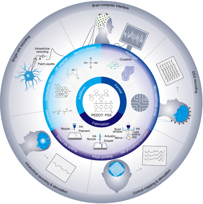
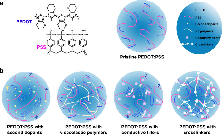
The growing demand for advanced neural interfaces that enable precise brain monitoring and modulation has catalyzed significant research into flexible, biocompatible, and highly conductive materials. PEDOT:PSS-based bioelectronic materials exhibit high conductivity, mechanical flexibility, and biocompatibility, making them particularly suitable for integration into neural devices for brain science research. These materials facilitate high-resolution neural activity monitoring and provide precise electrical stimulation across diverse modalities. This review comprehensively examines recent advances in the development of PEDOT:PSS-based bioelectrodes for brain monitoring and modulation, with a focus on strategies to enhance their conductivity, biocompatibility, and long-term stability. Furthermore, it highlights the integration of multifunctional neural interfaces that enable synchronous stimulation-recording architectures, hybrid electro-optical stimulation modalities, and multimodal brain activity monitoring. These integrations enable fundamentally advancing the precision and clinical translatability of brain-computer interfaces. By addressing critical challenges related to efficacy, integration, safety, and clinical translation, this review identifies key opportunities for advancing next-generation neural devices. The insights presented are vital for guiding future research directions in the field and fostering the development of cutting-edge bioelectronic technologies for neuroscience and clinical applications.


Conflict of interest statement

Conflict of interest: The authors declare no competing interests.

Figures

Fig. 1
Fig. 1
Schematic illustration showing the additives, fabrication and formats of PEDOT: PSS-based bioelectronics as well as their application in brain monitoring and modulation
Fig. 2
Fig. 2. Graphic illustration of PEDOT: PSS-based conductive materials.
a Schematic illustration of PEDOT:PSS. Left: molecular structure; Right: PEDOT:PSS in aqueous phase. b Schematic illustration of PEDOT:PSS with different additives in aqueous phase. Second dopants facilitate the PSS chain separation from the PEDOT:PSS complex (blue arrow) by charge screening effect. The phase separation enables PEDOT chain arrangement and the formation of π stacking (yellow dashed line), which improves the electrical transduction (yellow star). Viscoelastic polymers form hydrogen bond with PSS chain (blue dashed line), making the complex system more viscous and stretchable. Conductive fillers enhance the charge transport (white dashed line). Simultaneously, the fillers promote PSS removal (blue arrow) and PEDOT chain arrangement (yellow dashed line), all of which enhance the conductivity (yellow star). Crosslinkers form a long interconnected network with PEDOT:PSS systems by hydrogen bond with PSS (blue dashed line)
Fig. 3
Fig. 3. Different formats of PEDOT: PSS-based electrodes.
ac PEDOT:PSS-ionic liquid colloidal (PILC) ink for 3D-printed hydrogel. Copyright 2024, Springer Nature (Fig. S1). a: Images of a 3D array with a high aspect ratio (top) and high resolution (~50 µm) (bottom) fabricated via direct ink writing. b Impedance stability of PILC electronic devices incubated in PBS solution at 37 °C over 30 days. c Mechanical properties of printed PILC hydrogel, exhibiting a low Young’s modulus (750 kPa), comparable to biological tissues. df PEDOT:PSS/pHEMA film deposited on a rigid metal substrate (Au electrode) via electrochemical deposition for microelectrode array (MEA) fabrication. Copyright 2022, John Wiley and Sons (Fig. S2). d Images of the MEA shank (top) and a zoomed-in view of the pattern at the MEA tip (bottom). Gold: Au electrode; Black: PEDOT:PSS/pHEMA film. e Electrochemical impedance spectroscopy (EIS) measurement from 1 Hz to 100 kHz of bare gold MEAs (green), PEDOT/PSS (red), and PEDOT/PSS/pHEMA-coated (blue) MEAs. f EIS measurement of PEDOT/PSS (red) and PEDOT/PSS/pHEMA-coated (blue) during ageing test in PBS solution at 60 C for 7 days. gi: PEDOT:PSS film deposited on a flexible substrate (PET) via spin coating. Copyright 2023, John Wiley and Sons (Fig. S3). g Images of a transparent PEDOT:PSS-based ECoG grid wrapped around a cylindrical glass rod (5 mm diameter) (top) and a zoomed-in view of the electrode array (bottom). h EIS of the 30-channel ECoG grid. i Mechanical stability test of the ECoG grid, showing stable EIS values across 30 channels in four different states: original, kinked, folded, and sulcus
Fig. 4
Fig. 4. PEDOT:PSS-based bioelectronics for invasive, semi-invasive, and non-invasive brain modulation approaches.
PEDOT:PSS-based bioelectronics for invasive (ac), semi-invasive (df), and non-invasive (gi) brain modulation. ac PEDOT:PSS thin-film microelectrode arrays (MEAs) for brain stimulation and single-unit recording. Copyright 2022, The American Association for the Advancement of Science (Fig. S4). a The implantation process of a surface MEA (for surface stimulation) and a depth neuro-probe (for single-unit recording). Using a carrier glass pipette, the shank extension of the MEA was partially inserted into the brain, with ten electrodes reaching up to 500 μm below the pial surface. A micrograph of the MEA is shown (middle). b Illustration of the implanted MEA positioned both inside and on the brain surface, with PEDOT:PSS electrodes highlighted in blue. c Spike waveforms computed by averaging 50 traces recorded with the depth probe at 160 μm below the pial surface from a single neuron. df Block-Brush PEDOT:PSS film for ECoG recording. Copyright 2024, John Wiley and Sons (Fig. S5). d Schematic illustration of a 16-channel array implanted on the surface of the rat brain for barrel cortex recording under air-puff whisker stimulation. e Microscope image of the electrode array on the surface of the brain. The Block-Brush structure produced by SI-ATRP exhibited excellent conformity with the brain surface, whereas the electrode film fabricated by spin coating (SpinGA) presented an air gap. f Stimulation responses recorded from six individual low-impedance channels in the Block-Brush array. The dashed line indicates the timing of air-puff stimulation. gi PEDOT:PSS hydrogel-based electrode for EEG. Copyright 2023, Springer Nature (Fig. S6). g A photograph of a subject undergoing EEG monitoring. h Illustration of the EEG electrode. i EEG waveforms recorded using hydrogel, wet, and dry electrodes. The signals from the hydrogel electrode closely resembled those from the wet electrode but differed from those recorded with the dry electrode
Fig. 5
Fig. 5. PEDOT:PSS-based bioelectronics for multimodal brain modulation.
ac PEDOT:PSS-based microprobe system for optical stimulation and neurological activity sensing. Copyright 2020, Springer Nature (Fig. S7). a Schematic illustration of the microprobe system for optical stimulation and electrochemical sensing in deep brain tissue. b Position heat maps of animal activity, where hotter colors indicate longer duration at a given site. The map reveals that mice exhibit clear place preferences during the optogenetic stimulation period. c Spontaneous current spikes recorded during optogenetic stimulation. di A transparent electrode array based on PEDOT:PSS crosslinked with PEGDE and silk fibroin (P-PSF) for dual-modal neural-vascular activity probing. Copyright 2021, John Wiley and Sons (Fig. S8). d The transparent and conformable electrode was placed on the rat brain. e Time domain of neural electrical signals recorded from CH1-CH2, CH2-CH3, and CH3-CH4 showing changes during and shortly after photothrombosis (PT) stroke. f Schematic illustration of the transparent P-PSF electrodes for integrated vessel imaging and blood oxygen measurement under electrical stimulation. g Optical coherence tomography image of P-PSF electrodes (red dotted lines) positioned on the neurovascular system in the rat brain. h Relative changes in deoxy-Hb level in response to electrical stimulation induced by the P-PSF electrode compared to the referenced Au-SEBS electrode. i Statistical analysis of the time of arrival (ToA) and rise time upon stimulation

Similar articles

Cited by

References

    1. Kim, M., Lee, H., Nam, S., Kim, D.-H. & Cha, G. D. Soft bioelectronics using nanomaterials and nanostructures for neuroengineering. Acc. Chem. Res.57, 1633–1647 (2024). - PubMed
    1. Long, Y., Li, J., Yang, F., Wang, J. & Wang, X. Wearable and implantable electroceuticals for therapeutic electrostimulations. Adv. Sci.8, 2004023 (2021). - PMC - PubMed
    1. Koo, J. H., Song, J.-K., Kim, D.-H. & Son, D. Soft implantable bioelectronics. ACS Mater. Lett.3, 1528–1540 (2021).
    1. Li, J., Cao, J., Lu, B. & Gu, G. 3D-printed PEDOT:PSS for soft robotics. Nat. Rev. Mater.8, 604–622 (2023).
    1. Li, G. et al. Highly conducting and stretchable double-network hydrogel for soft bioelectronics. Adv. Mater.34, 2200261 (2022). - PubMed

LinkOut - more resources