. 2025 May 13;16(1):4414.
doi: 10.1038/s41467-025-59444-4.

ATP functions as a pathogen-associated molecular pattern to activate the E3 ubiquitin ligase RNF213

Affiliations

ATP functions as a pathogen-associated molecular pattern to activate the E3 ubiquitin ligase RNF213

Juraj Ahel et al. Nat Commun. .

Abstract

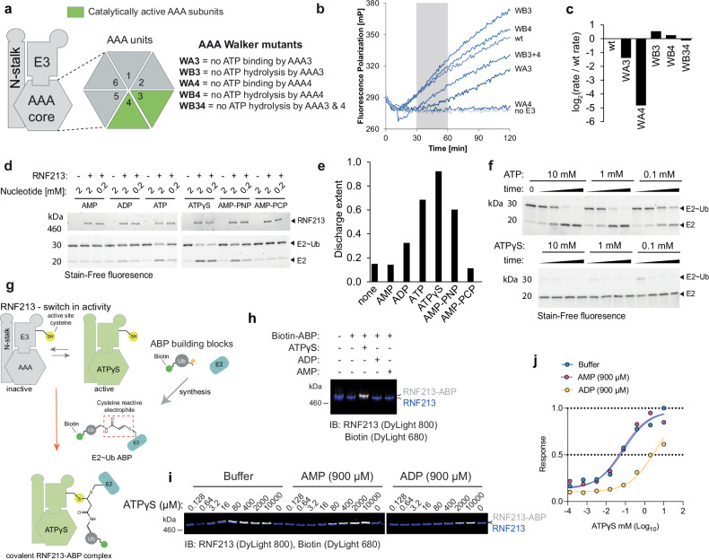
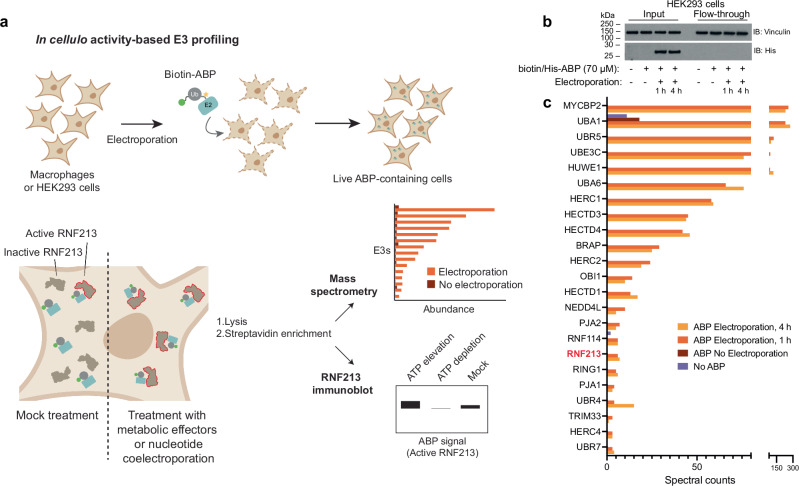
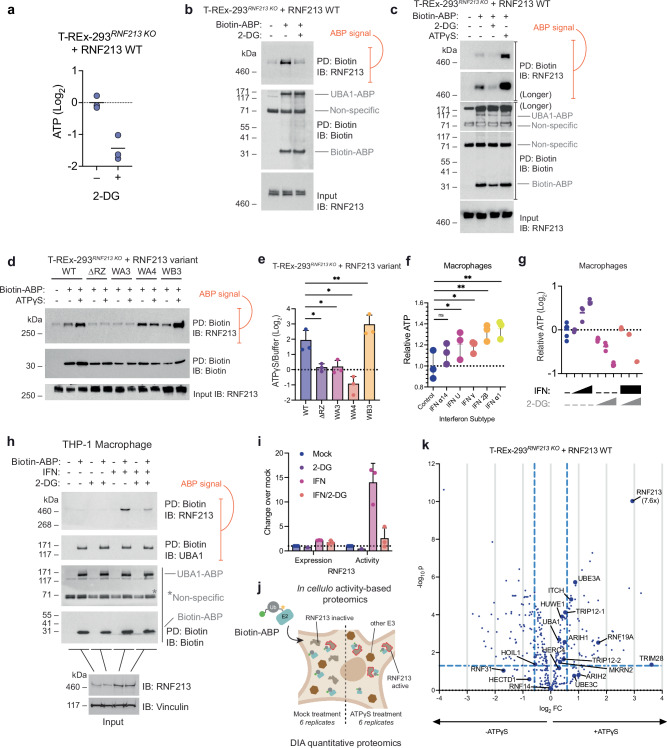
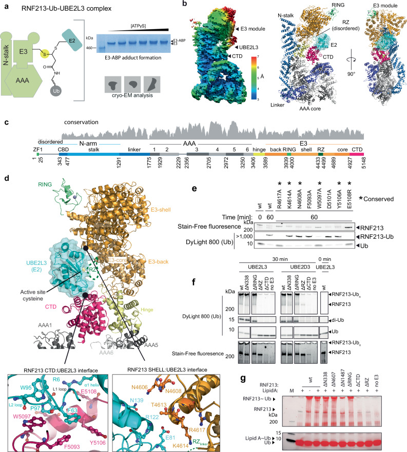
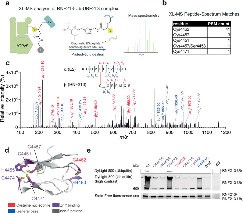
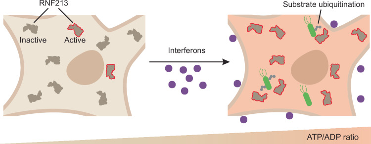
The giant E3 ubiquitin ligase RNF213 is a conserved component of mammalian cell-autonomous immunity, limiting the replication of bacteria, viruses and parasites. To understand how RNF213 reacts to these unrelated pathogens, we employ chemical and structural biology to find that ATP binding to its ATPases Associated with diverse cellular Activities (AAA) core activates its E3 function. We develop methodology for proteome-wide E3 activity profiling inside living cells, revealing that RNF213 undergoes a reversible switch in E3 activity in response to cellular ATP abundance. Interferon stimulation of macrophages raises intracellular ATP levels and primes RNF213 E3 activity, while glycolysis inhibition depletes ATP and downregulates E3 activity. These data imply that ATP bears hallmarks of a danger/pathogen associated molecular pattern, coordinating cell-autonomous defence. Furthermore, quantitative labelling of RNF213 with E3-activity probes enabled us to identify the catalytic cysteine required for substrate ubiquitination and obtain a cryo-EM structure of the RNF213-E2-ubiquitin conjugation enzyme transfer intermediate, illuminating an unannotated E2 docking site. Together, our data demonstrate that RNF213 represents a new class of ATP-dependent E3 enzyme, employing distinct catalytic and regulatory mechanisms adapted to its specialised role in the broad defence against intracellular pathogens.


Conflict of interest statement

Competing interests: S.V. is an author of a patent relating to the ABP technology and is also Founder and shareholder of Outrun Therapeutics. The remaining authors declare no competing interests.

Figures

Fig. 1
Fig. 1. Nucleotide-dependent regulation of RNF213 E3 activity.
a Schematic of RNF213 sub-modules (functional AAA3 and AAA4 in green) and mutations used to probe the role of the two functional AAA units in E3 ligase activity (WA3, K2387A; WA4, K2736A; WB3, E2449Q; WB4, E2806Q; WB34, E2449Q E2806Q; murine numbering). b Representative progress curves for the fluorescence polarization-based E3 activity assay. Reactions were carried out with wild type or mutant RNF213 in the presence of ubiquitin labeled sub-stoichiometrically with DyLight488. The time interval (30-60 min) used to determine rate constants is depicted in gray. c Rates of ubiquitin adduct formation (relative to wild type protein). d Discharge of Ub from purified UBE2L3~Ub discharge assay in the presence of RNF213 and various nucleotides. e Quantification of the discharge extent (free E2 / total E2). f Time course of UBE2L3~Ub discharge assay, using different nucleotide concentrations. g Schematic of the nucleotide-dependent activation of RNF213 HECT-like E3 activity, required to form a covalent complex with an activity-based probe (ABP, wavy bond corresponds to a triazole-ethyl linker). h Recombinant RNF213 was tested for transthiolation activity with a biotin-tagged ABP, in the presence or absence of 1 mM nucleotides indicated. Samples were resolved by SDS-PAGE and visualized by two-channel near-IR immunoblot using anti-RNF213 antibody (blue) and streptavidin (white). Panel representative of n = 3. i ABP analysis of ATPγS-dependent RNF213 activation in the presence of either ADP (900 μM) or AMP (900 μM). j Quantification of biotin signal determined in i. Mean values are plotted and non-linear fit curves shown (n = 2). Source data are provided as a Source Data file.
Fig. 2
Fig. 2. In cellula activity-based E3 ligase profiling.
a In cellula activity-based E3 ligase profiling involving the delivery of a biotin-tagged E2~Ub ABP into live cells by electroporation. Labeled E3s can be subsequently enriched and analysed by mass spectrometry or Western blot. The abundance of a recovered E3 is a proxy for its transthiolation activity, allowing relative changes to be inferred. b Western blot demonstrating intracellular delivery of ABP by electroporation followed by subsequent incubation for the specified time. Following lysis, the ABP and its labeled proteins can be subsequently captured with streptavidin resin. Panel shown representative of n = 3. c Mass spectrometry analysis showing total spectral counts from E3s and the two E1 enzymes (UBA1 and UBA6) enriched by Biotin-ABP electroporated into 293T cells. Plotted proteins had >10 times more spectral counts 1 h post-electroporation than ABP (based on UBE2D3) treatment without electroporation. E3s were selected based on Pfam domain terms. The E3 coverage was similar whether ABP was incubated for 1 or 4 h post-electroporation. Source data are provided as a Source Data file.
Fig. 3
Fig. 3. Assessment of RNF213 activation in live cells in response to changes in ATP levels.
a Treatment of RNF213 KO T-REx-293 cells stably overexpressing RNF213 (T-REx-293RNF213 KO + RNF213) with 30 mM 2-deoxyglucose (2-DG) for 3 h results in a 2-fold reduction in ATP levels. Cellular ATP levels were determined by luminescent ATP assay. Data are presented as mean values (n = 3). b In cellula ABP analysis of T-REx-293RNF213 KO + RNF213 cells that were mock, or 2-DG treated. RNF213 transthiolation activity is reduced in response to 2-DG treatment. UBA1, which also demonstrates transthiolation activity, is expressed at high levels and undergoes ABP labeling in a 2-DG insensitive manner. c Co-electroporation with ATPγS resulted in robust activation of RNF213 transthiolation activity. Panel representative of n = 2. d In cellula activity-based profiling of T-REx-293RNF213 KO cells stably overexpressing the stated RNF213 variant in response to ATPγS. e Quantification (densitometry) of fold increase in ABP labeling comparing ATPγS- to buffer-electroporated control, from d (biological replicates n = 3). Two-way ANOVA, P values compare the variants to WT (left to right); 0.0371 (*), 0.0391 (*), 0.0133 (*), 0.0038 (**); data are presented as mean values +/- SD (n = 3). f ATP levels in monocyte-derived macrophage THP-1 cells 16 h after treatment with 100 U/mL IFN ɑ14, Universal IFN (IFN-U), IFN γ, IFN 2β or IFN ɑ1. Cellular ATP levels were determined by luminescent ATP assay. Data are presented as mean values +/- SD. One-way ANOVA, F value = 6.915, Degrees of Freedom = 10; adj. P values (left to right); 0.1175 (ns), 0.0423 (*), 0.0492 (*), 0.0040 (**), 0.0025 (**); error bars are SD (n = 3, representative experiment comprising technical replicates shown). g Monocyte-derived macrophage THP-1 cells treated with 2-DG (3, 10, or 30 mM) and IFN ɑ1 (10, 100, or 1000 U/mL) for 16 h. Cellular ATP levels were determined by luminescent ATP assay. h In cellula transthiolation activity of endogenous RNF213 in IFN-stimulated macrophages that were mock or 2-DG treated. i Quantification (densitometry) of fold increase in Western band intensity across conditions, relative to mock treatment, from h (biological replicates n = 3; 2-DG-alone was included in 1 replicate as the signal was at or below detection threshold). Data are presented as mean values +/- SD. j Schematic illustrating how combining in cellula activity-based E3 ligase profiling with data-independent acquisition mass spectrometry (DIA-MS) allows the activity changes of a cohort of E3s to be simultaneously quantified upon cellular delivery of ATPγS. k Relative changes in ABP (based on UBE2L3) signal upon ATPγS delivery into T-REx-293RNF213 KO + RNF213 determined by in cellula activity-based E3 profiling and DIA-MS. Quantification was performed by analysing DIA spectra (n = 6) with DiaNN (v1.8.1) operating in library-free mode. Protein regulation was assessed using two-tailed differential expression analysis carried out in LIMMA using an empirical Bayes-moderated linear model and P values were corrected using Benjamini–Hochberg multiple hypothesis correction. The hashed vertical lines correspond to fold changes of 1/1.5 and 1.5, whereas the horizontal hashed line corresponds to an adjusted P-value cut-off of 0.05. Enlarged data points correspond to HECT/HECT-like E3s and the ubiquitin E1 UBA1. Two TRIP12 isoforms were detected and designated TRIP12-1 and TRIP12-2. Source data are provided as a Source Data file.
Fig. 4
Fig. 4. Cryo-EM structure of the RNF213-UBE2L3 transthiolation complex.
a Schematic of the covalent RNF213-UBE2L3~Ub complex, mimicking the transient E2-E3 transfer (transthiolation) intermediate. Inset illustrates complex formation by SDS-PAGE, demonstrating >90% labeling of full-length murine RNF213 (1.5 μM) at the highest ABP concentration (68 μM) in the presence of ATPγS (5 mM). Panel representative of n = 3. b Left, cryo-EM density of RNF213-UBE2L3~Ub complex at 3.5 Å resolution. A distinctive density appears between the E3 shell and the CTD domain of RNF213, into which UBE2L3 can be unambiguously docked. Right, ribbon representation of the RNF213-UBE2L3 model. Details about the cryo-EM reconstruction are given in Supplementary Fig. 4. c Domain architecture of RNF213. d Close-up view of the E3 module with bound UBE2L3 (color coded according to E3 portions, and UBE2L3 shown in light blue). The two interfaces with CTD and E3-shell are shown enlarged in insets. Comparison with E2 interfaces from other E3 enzymes is given in Supplementary Fig. 5. e Autoubiquitination assay used to validate the structurally determined UBE2L3 binding site. f Autoubiquitination assay with various deletion mutants of RNF213 when partnered with UBE2L3 or UBE2D3. Δ338 assesses the role of the N-terminal residues that were disordered in the cryo-EM structure. g Ubiquitination and lipid ubiquitination assays with RNF213 deletion mutants (auto-ubiquitination, top panel; lipid A ubiquitination, lower panel). Source data are provided as a Source Data file.
Fig. 5
Fig. 5. ABP mapping of the E3 cysteine nucleophile in the RZ domain.
a Cross-linking mass spectrometry (XL-MS) analysis of the full-length RNF213-ABP complex. The residue labeled by the ABP corresponds to the active site cysteine nucleophile (yellow circle), which was identified by XL-MS. b Cross-link peptide-spectrum matches (PSMs) that have an e-value lower than 1×10⁻⁴ with a false discovery rate (FDR) of <1% are tabulated. c Representative MS2 spectrum of a crosslinked peptide corresponding to Cys4462 in murine RNF213. The spectrum is for a z = 4+ precursor ion, observed precursor mass = 2369.186 Da; theoretical crosslinked peptide mass = 2369.189 Da. d Alphafold2 model of the murine RNF213 RZ domain. Consistent with ABP crosslinking analysis, UV-VIS absorbance spectroscopy, and activity assays, Cys4451, His4455, Cys4471, and Cys4474 coordinate a single metal ion. Cys4462 is the active site nucleophile essential for RNF213 transthiolation activity. His4483 is suitably positioned to serve as a general base that facilitates deprotonation of substrate nucleophiles. e Autoubiquitination assay with UBE2L3 using the point mutants of cysteine and histidine residues within the RZ domain. Source data are provided as a Source Data file.
Fig. 6
Fig. 6. Proposed model for RNF213 E3 activation upon sensing increased ATP/ADP ratio.
RNF213 E3 activity is tightly coupled to the cellular ATP/ADP ratio. Interferon stimulation causes an increase in ATP levels, which induces RNF213 E3 activity and substrate ubiquitination.

Similar articles

References

    1. Kato, H. et al. Differential roles of MDA5 and RIG-I helicases in the recognition of RNA viruses. Nature441, 101–105 (2006). - PubMed
    1. Chamaillard, M. et al. An essential role for NOD1 in host recognition of bacterial peptidoglycan containing diaminopimelic acid. Nat. Immunol.4, 702–707 (2003). - PubMed
    1. Morita, M. et al. Gene-targeted mice lacking the Trex1 (DNase III) 3’->5’ DNA exonuclease develop inflammatory myocarditis. Mol. Cell Biol.24, 6719–6727 (2004). - PMC - PubMed
    1. Idzko, M., Ferrari, D. & Eltzschig, H. K. Nucleotide signalling during inflammation. Nature509, 310–317 (2014). - PMC - PubMed
    1. Ernandes, M. J. & Kagan, J. C. Interferon-independent restriction of RNA virus entry and replication by a class of damage-associated molecular patterns. mBio12, 10.1128/mBio.00584-21 (2021). - PMC - PubMed

Substances

LinkOut - more resources