. 2025 May 13;16(1):4436.
doi: 10.1038/s41467-025-59630-4.

High-contrast in vivo fluorescence imaging exploiting wavelengths beyond 1880 nm

Affiliations

High-contrast in vivo fluorescence imaging exploiting wavelengths beyond 1880 nm

Jiayi Li et al. Nat Commun. .

Abstract

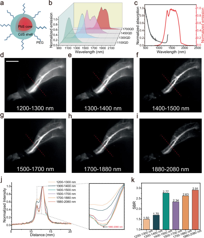
The second near-infrared (NIR-II) window is widely acknowledged for its excellent potential in in vivo fluorescence imaging. Currently, NIR-II fluorescence imaging predominantly operates within the 900-1880 nm spectral range, while the region beyond 1880 nm has been disregarded due to the large light absorption of water. Based on a refined understanding of the effect of light absorption on imaging, we propose an approach that utilizes the previously neglected region surrounding the water absorption peak at ~1930 nm for imaging. Both simulations and experiments confirm that the water absorption contributes positively to imaging, enabling high-contrast in vivo fluorescence imaging in the 1880-2080 nm window. To further assess the applicability of this approach in different biological media, we extend our focus to fluorescence imaging in adipose tissue. This leads to the expansion of the imaging window to 1700-2080 nm, owing to the unique light absorption characteristics of adipose tissue. Our results demonstrate that the 1700-2080 nm region provides optimal imaging quality in adipose tissue, attributing to its moderate absorption and low scattering. This work advances our understanding of the interplay between light absorption and photon scattering in bioimaging, providing an insight for selecting optimal imaging windows to achieve high-contrast fluorescence imaging.


Conflict of interest statement

Competing interests: The authors declare no competing interests.

Figures

Fig. 1
Fig. 1. The schematic diagram of photon propagation in tissue and the simulation results of bioimaging via the Monte Carlo method.
a Schematic of photon propagation in biological tissues with high and low light absorption, and the corresponding imaging effects. b The absorption spectrum of water within 700–2300 nm. c Simulated images within 1200–1300, 1300–1400, 1400-1500, 1500–1700, 1700–1880, and 1880–2080 nm, through routine biological tissue of 1 mm thickness, where the absorption coefficient of water was considered as the tissue absorption coefficient and the setting of tissue scattering coefficient could be found in the “Methods” section. d SBR analysis of the simulation results. n = 3 positions were randomly selected for analysis, data are presented as mean ± SD. e The structure similarity index measure (SSIM) analysis of the simulation results.
Fig. 2
Fig. 2. Comparison of in vivo fluorescence imaging of mice within various NIR bands.
a Schematic design of the PEGylated PbS/CdS core-shell QDs. b Normalized emission spectra of four kinds of PEGylated QDs in aqueous dispersion. c Normalized absorption and emission spectra of four kinds of PEGylated QDs mixed in a certain ratio in water. The hind limb fluorescence images of the same mouse in d 1200–1300 nm, e 1300–1400 nm, f 1400–1500 nm, g 1500–1700 nm, h 1700–1880 nm, and i 1880–2080 nm. Scale bars: 10 mm. j Cross-sectional fluorescence intensity profiles along the red dashed lines of the vessel in (di) and the localized magnification of the background signal. k SBR analysis of the imaging results in (di).
Fig. 3
Fig. 3. Comparison of in vivo NIR-II fluorescence imaging upon a bright background and multi-channel imaging of mice.
a Simulated images in 1300–1400, 1400–1500, 1500–1700, 1700–1880 and 1880–2080 nm of the line source above a rectangular background (depth = 1.3 mm) through a routine biological tissue of 1 mm thickness, where the absorption coefficient of water was considered as the tissue absorption coefficient and the setting of tissue scattering coefficient could be found in the “Methods” section. b Fluorescence intensity profiles on the cross-section of the simulated samples above the rectangular backgrounds. c SBR analysis of the simulation results in (a). n = 3 positions were randomly selected for analysis, data are presented as mean ± SD. d SSIM analysis of the simulation results in (a). Whole-body fluorescence imaging of the same mouse in e 1300–1400 nm, f 1400–1500 nm, g 1500–1700 nm, h 1700–1880 nm and i 1880–2080 nm bands. Scale bars: 10 mm. j Cross-sectional fluorescence intensity profiles along the red dashed lines of the blood vessels in (ei). k SBR analysis of the imaging results in (ei). l The perception-based image quality evaluator (PIQE) analysis of the liver areas highlighted by the blue boxes in (ei). m Fluorescence images of the centrifuge tubes containing 1450QD and 1700QD in 1400–1500 and 1880–2080 nm bands. np In vivo dual-channel fluorescence imaging of mice. n An abdominal image of mouse intraperitoneally injected with 1450QD. o A vascular image of mouse intravenously injected with 1700QD. p The merged image of (n, o). Scale bars: 10 mm.
Fig. 4
Fig. 4. The simulation results of NIR fluorescence imaging through adipose tissue via the Monte Carlo method.
a Reduced scattering spectra of adipose tissue within 600–2200 nm and its fitted curve. b Absorption spectra of adipose tissue within 600–2200 nm. c Simulated fluorescence images of a line source passing through the adipose tissue of 1, 2 and 3 mm thicknesses within the 900–1100, 1100–1300, 1300–1400, 1400–1500, 1500–1700, and 1700–2080 nm windows. d SBR analysis of the simulation results at different depths. n = 3 positions were randomly selected for analysis, data are presented as mean ± SD. e SSIM analysis of the simulation results at different depths.
Fig. 5
Fig. 5. In vitro and in vivo experiments for NIR fluorescence imaging through adipose tissue.
a A bright-field image showing two capillaries loaded with hybrid QDs fluorescent probes and buried in the porcine adipose tissue. b Fluorescence images of capillaries through adipose tissue of 2 mm thickness within 900–1100, 1100–1300, 1300–1400, 1400–1500, 1500–1700, and 1700–2080 nm windows. c Fluorescence images of capillaries through adipose tissue of 1, 2, and 3 mm thickness at the 1700–2080 nm window. d Normalized fluorescence intensity profiles along the red dashed lines in (b). e SBR analysis of the profiles in (d). f FWHM analysis of the profiles in (d). g, h Fluorescence images of rabbit bile ducts without adipose tissue covered in the 1700–2080 nm window. Scale bars of (g): 5 mm. CHD common hepatic duct, CBD common bile duct. i Normalized fluorescence intensity profile along the yellow dashed line in (g) and the corresponding Gaussian-fitted FWHM value. j, k Fluorescence images of rabbit bile ducts covered with porcine adipose tissue of ~2 mm thickness in the 1700–2080 nm window. Scale bars of (j): 5 mm. l Normalized fluorescence intensity profile along the yellow dashed line in (j) and the corresponding Gaussian-fitted FWHM value, where the dots represent the raw data and the blue line represents Gaussian fitted curves.

Similar articles

References

    1. Nagaya, T., Nakamura, Y. A., Choyke, P. L. & Kobayashi, H. Fluorescence-guided surgery. Front. Oncol.7, 314 (2017). - PMC - PubMed
    1. Welsher, K. et al. A route to brightly fluorescent carbon nanotubes for near-infrared imaging in mice. Nat. Nanotechnol.4, 773–780 (2009). - PMC - PubMed
    1. Hong, G. et al. Through-skull fluorescence imaging of the brain in a new near-infrared window. Nat. Photonics8, 723–730 (2014). - PMC - PubMed
    1. Wang, P. et al. NIR-II nanoprobes in-vivo assembly to improve image-guided surgery for metastatic ovarian cancer. Nat. Commun.9, 2898 (2018). - PMC - PubMed
    1. Hu, Z. et al. First-in-human liver-tumour surgery guided by multispectral fluorescence imaging in the visible and near-infrared-I/II windows. Nat. Biomed. Eng.4, 259–271 (2020). - PubMed

LinkOut - more resources