. 2025 May 13;11(1):77.
doi: 10.1038/s41522-025-00710-0.

SadB, a mediator of AmrZ proteolysis and biofilm development in Pseudomonas aeruginosa

Affiliations

SadB, a mediator of AmrZ proteolysis and biofilm development in Pseudomonas aeruginosa

Yossi Ben-David et al. NPJ Biofilms Microbiomes. .

Abstract

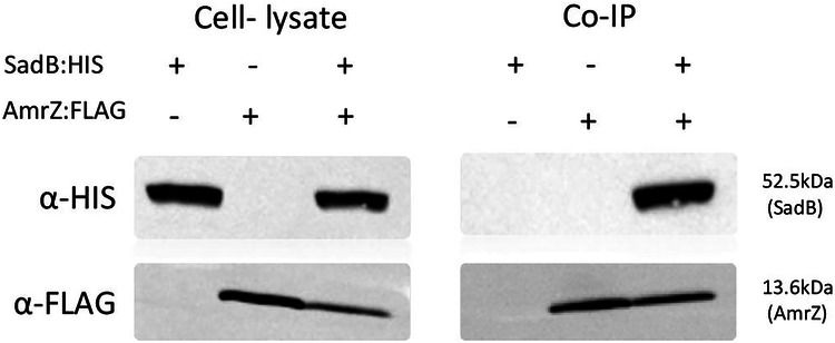
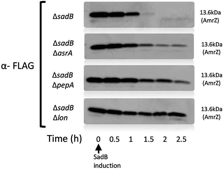
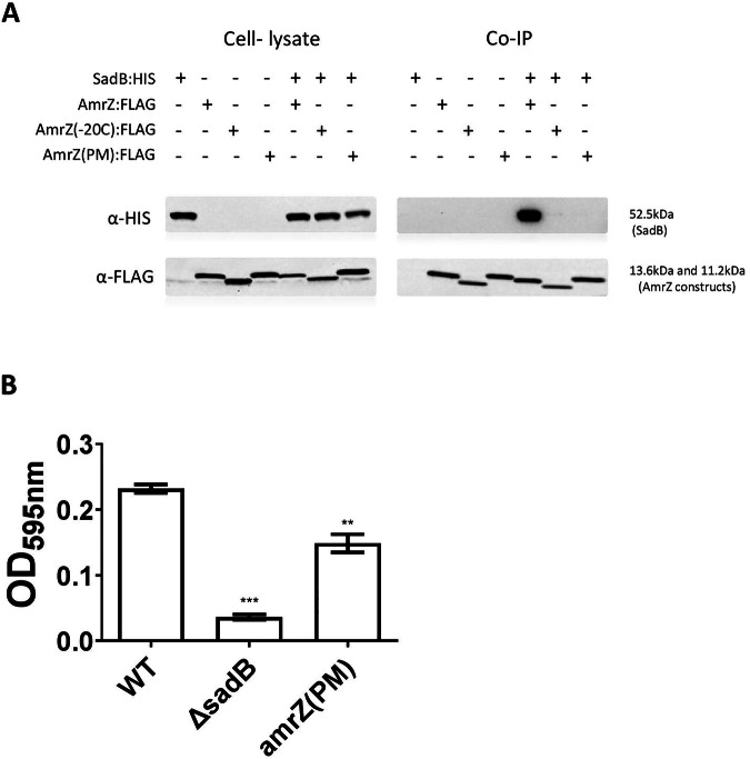
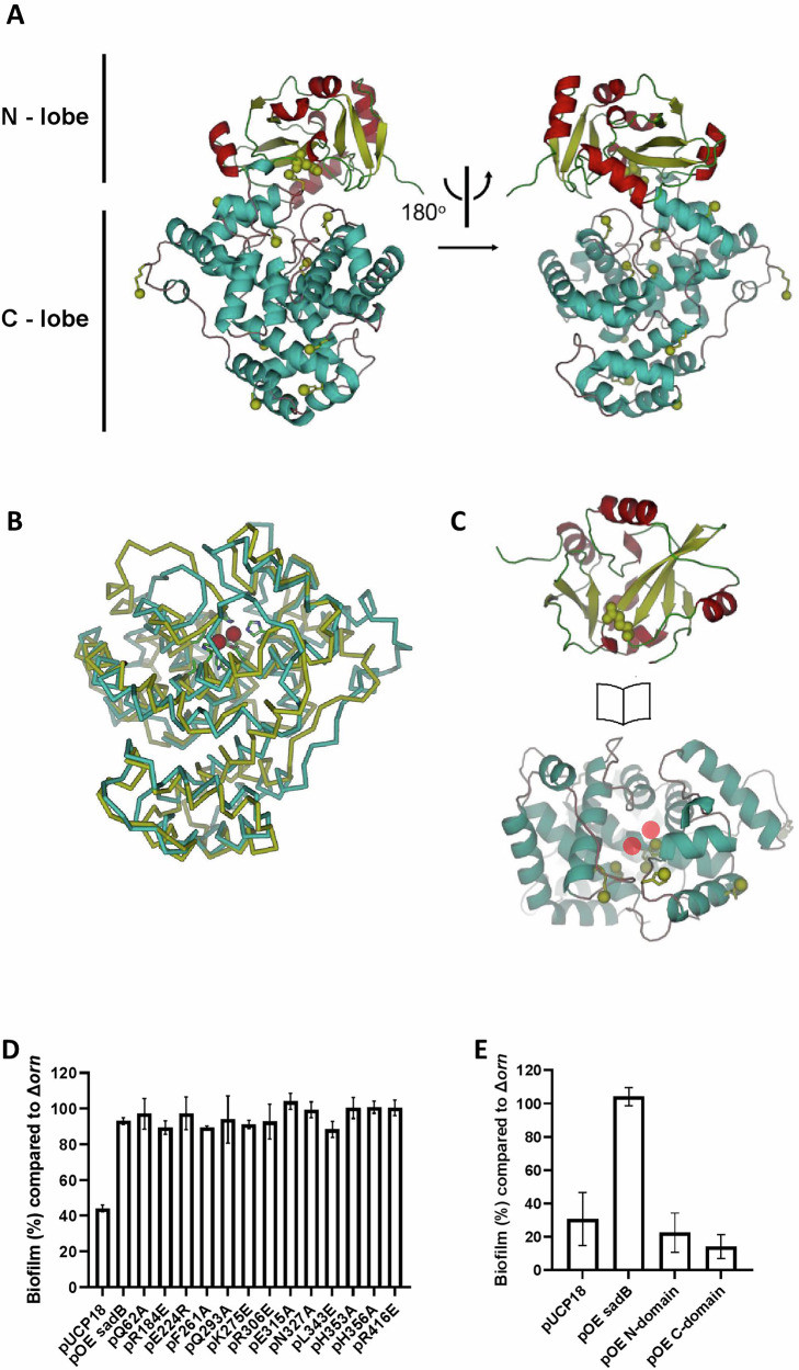
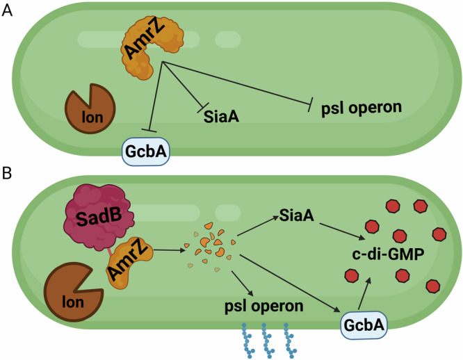
The ability of bacteria to commit to surface colonization and biofilm formation is a highly regulated process. In this study, we characterized the activity and structure of SadB, initially identified as a key regulator in the transition from reversible to irreversible surface attachment. Our results show that SadB acts as an adaptor protein that tightly regulates the master regulator AmrZ at the post-translational level. SadB directly binds to the C-terminal domain of AmrZ, leading to its rapid degradation, primarily by the Lon protease. Structural analysis suggests that SadB does not directly interact with small molecules upon signal transduction, differing from previous findings in Pseudomonas fluorescens. Instead, the SadB structure supports its role in mediating protein-protein interactions, establishing it as a major checkpoint for biofilm commitment.


Conflict of interest statement

Competing interests: The authors declare no competing interests.

Figures

Fig. 1
Fig. 1. AmrZ and ProE levels are post-translationally regulated by SadB.
A Scatter plot of a Spearman correlation of proteomics and transcriptomics results of a sadB mutant (ΔsadB) strain, as compared to the WT strain, presented as log of fold-change. Spearman’s rho was 0.603 and the p value was <0.00001. B Western blot exhibiting FLAG-conjugated AmrZ and ProE expression levels in a sadB over-expressing strain (pOE sadB) and in a sadB mutant strain carrying an empty vector (ΔsadB/pUCP18), as compared to the WT strain carrying an empty vector (PAO1/pUCP18). The image is representative of three independent repeats. C Real-time PCR analysis measuring amrZ and proE mRNA levels in a sadB over-expressing strain (pOE sadB) and a sadB mutant strain transformed with an empty vector (ΔsadB/pUCP18), as compared to the WT strain also transformed with an empty vector (PAO1/pUCP18).
Fig. 2
Fig. 2. AmrZ and ProE mutants abolish ΔsadB-related phenotypes.
A Swarming assay. B Static biofilm assay. C Flow cell assay and imaris biovolume quantification of the flow cell results. D Cellular c-di-GMP measurements for the WT, sadB mutant (ΔsadB), amrZ mutant (ΔamrZ), proE mutant (ΔproE), sadB amrZ double mutant (ΔsadB ΔamrZ), sadB proE double mutant (ΔsadB ΔproE) and the sadB amrZ proE triple mutant (ΔsadB ΔamrZ ΔproE) strains. The static biofilm assay reflects the average of three independent repeats, with each experiment involving six replicates. Error bars represent standard deviation. For the swarming assay, a representative plate of each strain from three replicates in three independent experiments is shown. For the flow cell assay, one representative image of each strain of three independent experiments is shown. In cellular c-di-GMP measurements, each bar represents the average of three independent experiments. * Indicates a statistically significant result where P < 0.05, ** indicates a statistically significant result where P < 0.01 and *** indicates a statistically significant result where P < 0.001.
Fig. 3
Fig. 3. SadB interacts with AmrZ at the protein level.
Co-Immunoprecipitation with anti-FLAG resin testing for interactions between co-expressed polyhistidine-conjugated SadB and FLAG-conjugated AmrZ using the sadB:HIS and amrZ:FLAG. Cell lysates from SadB- (sadB:HIS) and AmrZ-expressing cells (amrZ:FLAG) served as negative controls. The blot shown is representative of three independent repeats.
Fig. 4
Fig. 4. SadB promotes AmrZ degradation through proteolysis, primarily by the quality control protease Lon.
A degradation assay of FLAG-conjugated AmrZ over time after SadB appearance (∆sadB/ pRBSmod sadB:HIS and pOE amrZ:FLAG) in mutant strains deleted of different protease (∆sadBasrA, ∆sadBpepA, ∆sadBlon). Each blot is a representative of 3 independent repeats.
Fig. 5
Fig. 5. The degradation process depends on a conserved amino acid residue located near the C-terminal end of AmrZ.
A ConSurf results of AmrZ, presenting conservation/variability of amino acids between numerous Pseudomonas species. The blue square highlights the conserved region within the variable sequence. B A diagram presenting different amrZ constructs, namely, complete amrZ and amrZ lacking the sequence encoding the C-terminal ten amino acids (amrZ(-10C):FLAG) or the last twenty residues (amrZ(-20C):FLAG). C A degradation assay of the different AmrZ:FLAG constructs (pOE amrZ:FLAG, pamrZ(-10C):FLAG, pamrZ(-20C):FLAG) over time following SadB appearance (∆sadB/pRBSmod sadB:HIS). Each blot is a representative of three independent repeats. D A diagram presenting the three C-terminal domain point mutations (PM) introduced into amrZ (pamrZ(PM):FLAG), encoding Q91A, A94Q or L95Q. E A degradation assay of the different AmrZ:FLAG constructs (pOEamrZ:FLAG, pamrZ(PM):FLAG) following SadB appearance (∆sadB/pRBSmod sadB:HIS). Each blot is a representative of three independent repeats.
Fig. 6
Fig. 6. The conserved C-terminal sequence is crucial for SadB binding and regulation.
A Co-Immunoprecipitation with anti-FLAG resin testing for interaction between co-expressed Poly histidine-conjugated SadB and different constructs of FLAG-conjugated AmrZ (sadB:HIS and amrZ:FLAG; amrZ(-20C):FLAG; amrZ(PM):FLAG). Cell lysates from SadB (sadB:HIS) and AmrZ-expressing cells (amrZ:FLAG, amrZ(-20C):FLAG and amrZ(PM):FLAG) served as negative controls. The blot is representative of three independent repeats. B static biofilm assay comparing the WT strain (WT), a sadB mutant (ΔsadB) and a 3 amino acid point-mutated AmrZ variant that is resistant to SadB binding and subsequent degradation (amrZ(PM)). An average of 3 independent experiments is presented; each experiment contains six replicates. Error bars represent the standard deviation. ** indicates a statistically significant result where P < 0.01 and *** indicates a statistically significant result where P < 0.001.
Fig. 7
Fig. 7. Biofilm modulation by SadB does not require ligand binding to the HDOD pocket, although both the N– and C-domains are necessary.
A Domain organization of SadB, with the N-domain colored red and yellow, and the C-domain cyan. Note the groove between the two domains. Sites modified by site-directed mutagenesis performed in this study are highlighted by green spheres. B Super-imposition of the SadB C-domain (cyan) and the HDOD domain from G. sulfurreducens (PDB entry 3HC1) with two iron ions bound in the HDOD pocket (red spheres). While iron coordination in the G. sulfurreducens structure is mediated by five histidine residues and one aspartate residue, the corresponding sites in SadB lack three histidine residues. C “Open book” representation of the separated N- and C-domains, revealing the inter-domain interacting surfaces. D&E. Static biofilm assay, with the genetic background of all the tested strains being ΔsadBΔorn. All SadB modifications are indicated in Supplementary Fig. 4. D Examining the effects of 13 different point mutations on SadB behavior. E Testing the behavior of the SadB N- and C-domains on their own. The average of three independent repeats is shown, with each repeat comprising six replicates. Error bars represent standard deviation.
Fig. 8
Fig. 8. Scheme summarizing SadB effect on P. aeruginosa.
A Inactive/absent SadB allows AmrZ to accumulate in the cell leading to repression of biofilm formation. B Higher levels of SadB mark AmrZ for degradation inducing c-di-GMP accumulation, PSL production and subsequent biofilm formation.

References

    1. Hsueh, P. R. et al. Persistence of a multidrug-resistant Pseudomonas aeruginosa clone in an intensive care burn unit. J. Clin. Microbiol36, 1347–1351 (1998). - PMC - PubMed
    1. Aloush, V., Navon-venezia, S., Seigman-igra, Y., Cabili, S. & Carmeli, Y. Multidrug-resistant Pseudomonas aeruginosa: risk factors and clinical impact. Society50, 43–48 (2006). - PMC - PubMed
    1. Kwon, S., Schweizer, M. L. & Perencevich, E. N. National Institute of Allergy and Infectious Disease (NIAID) funding for studies of hospital-associated bacterial pathogens: are funds proportionate to burden of disease?. Antimicrob. Resist Infect. Control1, 5 (2012). - PMC - PubMed
    1. Hirsch, E. B. & Tam, V. H. Impact of multidrug-resistant Pseudomonas aeruginosa infection on patient outcomes. Expert Rev. Pharmacoecon Outcomes Res.10, 1–18 (2011). - PMC - PubMed
    1. Hill, D. et al. Antibiotic susceptibility of Pseudomonas aeruginosa isolates derived from patients with cystic fibrosis under aerobic, anaerobic, and biofilm conditions. J. Clin. Microbiol43, 5085–5090 (2005). - PMC - PubMed

MeSH terms

Substances

LinkOut - more resources