. 2025 May 13;15(1):16586.
doi: 10.1038/s41598-025-01707-7.

Multi-wavelength imaging photoplethysmography for non-invasive and non-contact assessment of burn severity

Affiliations

Multi-wavelength imaging photoplethysmography for non-invasive and non-contact assessment of burn severity

You-Rim Park et al. Sci Rep. .

Abstract

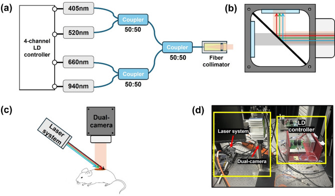
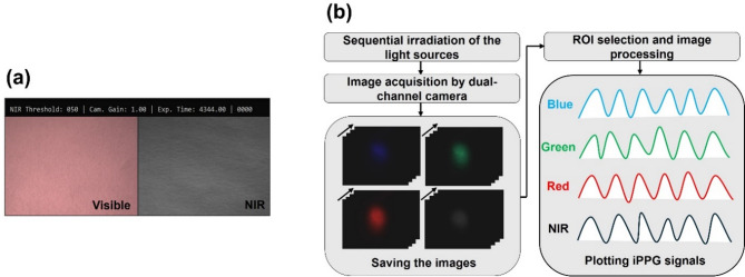
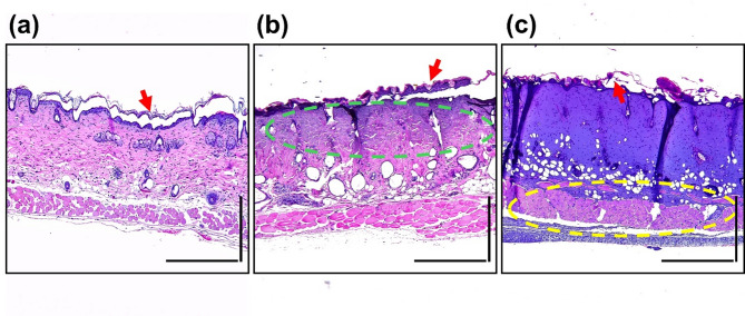
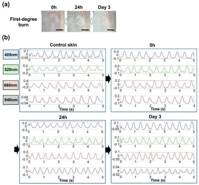
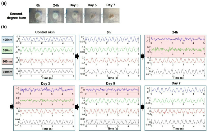
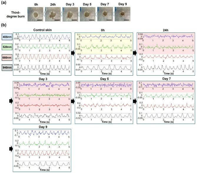
We report a non-contact burn severity assessment system using the image-based photoplethysmography (IPPG) technique by fabricating a multi-wavelength imaging system. In this burn assessment system, four wavelengths (visible light wavelengths of 405 nm, 520 nm, 660 nm, and near-infrared wavelength of 940 nm) were used, and burn severity was identified based on the fact that each wavelength has different penetration depths. Each wavelength was set to irradiate with the same optical power (1 mW/cm²), and IPPG was acquired using images captured at 35 frames per second for wavelengths with different penetration depths. To measure burn severity, we created burn lesion models using hairless mice. For each degree of burn, we acquired images of the burn area at four different wavelengths, measured IPPG from the acquired images, and observed the signal change at each wavelength to evaluate burn severity. In addition, while monitoring the healing process, we observed that IPPG recovered as the blood flow in the tissue normalized. Through the results of this study, we expect that IPPG technology will be used not only as a non-contact technology to evaluate burn severity, but also as a new method to monitor the burn recovery process in real time.

Keywords: Burn depth; Dual-camera; Imaging photoplethysmography; Laser diodes; Multi-wavelength.


Conflict of interest statement

Declarations. Competing interests: The authors declare no competing interests.

Figures

Fig. 1
Fig. 1
Light source and camera system for burn depth assessment (a) The schematic diagram of the laser system combining multiple wavelengths (b) The schematic diagram of a dual-camera accommodating visible and near-infrared light (c) The schematic diagram of the imaging system (d) Experimental system setup.
Fig. 2
Fig. 2
The process of acquiring IPPG signals at each wavelength (a) Custom-designed graphical user interface (GUI) displaying visible and near-infrared images on a split screen, with a field of view (FOV) of 4.8 × 3.65 cm². (b) Block diagram including the process of acquiring IPPG signals through multiple wavelengths imaging in burn lesion models.
Fig. 3
Fig. 3
Histological images according to burn lesion classification (a) First-degree burn; the red arrow indicates partial desquamation of the stratum corneum. (b) Second-degree burn; the green dashed circle indicates the disruption of the basketweave arrangement of collagen fibers and their abnormal thickening due to dermal damage. (c) Third-degree burn; the yellow dashed circle indicates the loss of the polygonal arrangement of muscle fibers. Scale bar = 500 µm.
Fig. 4
Fig. 4
(a) Captured images showing the progression of wound healing in the mouse model with the first-degree burn for 3 days. Scale bar = 0.5 cm. (b) IPPG signals at different wavelengths in the burn area of the first-degree burn over 3 days. In the first-degree burn, it was confirmed that IPPG signals were not damaged at any wavelength.
Fig. 5
Fig. 5
(a) Captured images showing the progression of wound healing in the mouse model with the second-degree burn for 7 days. Scale bar = 0.5 cm. (b) IPPG signals at different wavelengths in the burn area of second-degree burn over 7 days. The transparent red boxes indicate the damaged IPPG signals. 24 h after the burn injury, IPPG signals at the blue and green wavelengths were damaged. On the seventh day, IPPG signals were restored at all wavelengths, confirming the restoration of the blood vessels.
Fig. 6
Fig. 6
(a) Captured images showing the progression of wound healing in the mouse model with the third-degree burn for 9 days. Scale bar = 0.5 cm. (b) IPPG signals at different wavelengths in the burn area of the third-degree burn over 9 days. The transparent yellow boxes indicate weakened IPPG signals and transparent red boxes indicate damaged IPPG signals. The maximum damage to IPPG signals, including those at the blue, green, and red wavelengths, was observed on the third day. On the nineth day, IPPG signals were restored at all wavelengths, confirming the restoration of the blood vessels.

Similar articles

References

    1. Evers, L. H., Bhavsar, D. & Mailänder, P. The biology of burn injury. Exp. Dermatol.19, 777–783. 10.1111/j.1600-0625.2010.01105.x (2010). - PubMed
    1. Çakir, B. & Yeǧen, B. Ç. Systemic responses to burn injury. Turk. J. Med. Sci.34, 215–226. 10.1097/00005373-197010000-00008 (2004).
    1. Lateef, Z. et al. The cutaneous inflammatory response to thermal burn injury in a murine model. Int. J. Mol. Sci.20, 538. 10.3390/ijms20030538 (2019). - PMC - PubMed
    1. Lu, S. Burn wound healing. Chin. Burn Surg. 207–248. 10.1007/978-94-017-85754_9 (2015).
    1. Warby, R. & Maani, C. V. Burns Classification. StatPearlshttps://www.ncbi.nlm.nih.gov/books/NBK539773/ (2023). - PubMed

LinkOut - more resources