. 2025 May 13;15(1):16595.
doi: 10.1038/s41598-025-99187-2.

Edaravone dexborneol provides neuroprotective benefits by suppressing ferroptosis in experimental intracerebral hemorrhage

Affiliations

Edaravone dexborneol provides neuroprotective benefits by suppressing ferroptosis in experimental intracerebral hemorrhage

Han Li et al. Sci Rep. .

Abstract

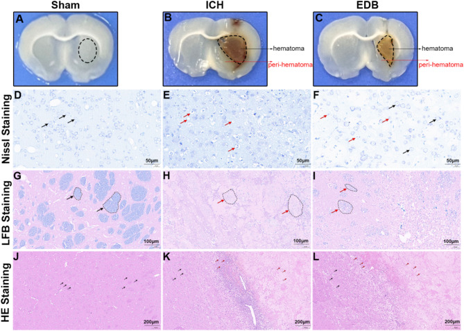
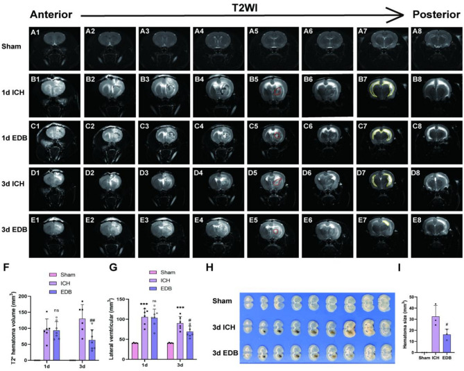
Edaravone dexborneol (EDB) is widely recognized for its anti-inflammatory and antioxidant properties and is clinically applied in the treatment of acute cerebral infarction. Ferroptosis is a critical process in the pathophysiology of brain injury following intracerebral hemorrhage (ICH). However, it remains unclear whether EDB can ameliorate ICH through the modulation of ferroptosis. This study aimed to evaluate the function and mechanism of EDB in treatment of ICH. With a male rat ICH model, animal behavior tests, histopathological staining, magnetic resonance imaging and evans blue staining were used to evaluate the neural protective function of EDB on ICH rats. The potential molecular mechanism was investigated using RNA sequencing. With the administration of Fer-1, a range of ferroptosis-related biomarkers, including Fe2+, 4-hydroxynonenal, malondialdehyde, etc., were analyzed to ascertain whether EDB confers neuroprotective effects through the modulation of P53/GPX4 pathways to inhibit ferroptosis. Finally, the findings were further corroborated using an in vitro ICH model with a P53 inhibitor. EDB has the potential to markedly enhance nerve and motor function, mitigate pathological damage, facilitate hematoma clearance, and repair BBB injury in ICH rats. KEGG analysis revealed that the differentially expressed genes were associated with signaling pathways, including P53 and ferroptosis. Both EDB and Fer-1 substantially reduced the concentrations of Fe2+, 4-hydroxynonenal, malondialdehyde, increased the amount of anti-oxidants, decreased the expression of P53, and concurrently upregulated the expression of GPX4. Besides, the P53 inhibitor PFT-α was observed to significantly reduce the levels of 4-HNE and lipid peroxides, while concurrently increasing the expression of GPX4. This investigation has shed light on the crucial neuroprotective role of EDB by regulating ferroptosis in ICH disease, which provided a theoretical basis for the clinical application of EDB in the treatment of ICH.

Keywords: Edaravone dexborneol; Ferroptosis; GPX4; Intracerebral hemorrhage; P53.


Conflict of interest statement

Declarations. Competing interests: The authors declare no competing interests.

Figures

Fig. 1
Fig. 1
Experimental design and animal behavior test. (A,B) Schematic diagram of the in vivo experimental procedure. (C) mNSS results assessed neurological impairment of rats. (D) Rotarod test results showed the duration time of rats staying on the rod. (E) The corner turn test show frequency of right turns in rats. ##P < 0.01 vs. ICH group; ###P < 0.001 vs. ICH group. N = 10 in Sham group; N = 15 in ICH and EDB group.
Fig. 2
Fig. 2
Detection of pathological injury and inflammation infiltration by Nissl, LFB and HE staining. (AC) Representative whole brain slices of different groups with 1 mm thickness. The striatal area demarcated by black oval outlines in (A) was used for Nissl staining, HE staining, and LFB staining; The peri-hematomal regions (0.5 mm from the hematoma border) indicated by arrows in (B,C) were selected for Nissl and HE staining, while the core hematoma areas outlined by black ovals were utilized for LFB staining. (DF) Representative Nissl staining images in the perihematomal region of different groups. Black arrow showed normal neurons, while red arrow showed abnormal neurons. (GI) Representative LFB staining images in the hematomal region of different groups. Black arrow showed normal myelin plaque, while red arrow showed abnormal myelin plaque. (JL) Representative HE staining images in the perihematomal region of different groups. Black arrow showed healthy neurons, while red arrow showed infiltrated inflammatory cells. Scale bar is 50 μm in (DF), 100 μm in (GI), and 200 μm in (JL).
Fig. 3
Fig. 3
Detection of hematoma volume and lateral ventricle volume by MRI and tissue section. (AE) T2-weighted MRI scans (coronal plane) showing hematoma changes at 1 day and 3 days after ICH. Red dashed lines indicate hematoma size and yellow blood lines indicate lateral ventricle volume. (F) Quantitative analysis of hematoma volume on T2WI images at 1 day and 3 days after ICH. (G) Quantitative analysis of lateral ventricle volume on T2WI images at 1 day and 3 days after ICH. All data were expressed as mean ± SD. (H) Macroscopic images of rat brains (0.5 mm thick) obtained by dissection. (I) Quantitative analysis of hematoma volume in brain tissue sections. All data were expressed as mean ± SD. ***P < 0.001 vs. Sham group; nsP > 0.05 vs. ICH group; #P < 0.05 vs. ICH group; ##P < 0.01 vs. ICH group.
Fig. 4
Fig. 4
Detection of BBB integrity by Evans blue staining and WB. (A) Representative images showing Evans blue extravasation 3 days after ICH induction. (B) Representative western blot images of occludin, ZO-1 and MMP-9 in striatum brain tissue of rat brain. Occludin belt was cropped between 50 and 70 kDa. ZO-1 belt was cropped between 150 and 250 kDa. MMP-9 belt was cropped between 70 and 100 kDa. GAPDH belt was cropped between 35 and 40 kDa. ZO-1 and GAPDH were cropped from the same gel, while Occludin and MMP-9 were cropped from different gels. Full-length blots were presented in Additional file 1: Fig. S1A–D. (C) Quantification of Evans blue content in the right brain hemisphere of rats. (DF) Quantification of western blot results of occludin, ZO-1 and MMP-9. **P < 0.01 vs. Sham group; ***P < 0.001 vs. Sham group; ##P < 0.01 vs. ICH group; ###P < 0.001 vs. ICH group. All data were expressed as mean ± SD of 5 rats.
Fig. 5
Fig. 5
Differential expression genes and pathway enrichment analysis. (A) Volcano plot displayed DEGs between Sham and ICH. (B) Volcano plot displayed DEGs between ICH and EDB. (C) Venn diagram showed the common DEGs between Sham vs. ICH and ICH vs. EDB. (D) Heatmap demonstrated the 684 unique genes. (E) Gene ontology biological processes (GO-BP), cellular component (GO-CC) and molecular functions (GO-MF). (F) KEGG enrichment analysis showed signaling pathway that the DEGs involved in.
Fig. 6
Fig. 6
Detection of ferroptosis-related markers. (AC) Brain sections were subjected to Prussian blue staining to detect the deposition of iron in the perihematoma of brain tissues, Bar = 200 μm. (DH) The content of Fe2+, 4-HNE, MDA, GSH, and T-SOD in the striatum tissue of rats were detected. (I) Representative western blot images of P53, GPX4, NRF2, SLC7A11, HIF1α, and HMOX1 at day 3 and day 7. P53 belt was cropped between 50 and 70 kDa. GPX4 belt was cropped between 17 and 25 kDa. NRF2 belt was cropped between 150 and 250 kDa. SLC7A11 belt was cropped between 40 and 50 kDa. HIF1α belt was cropped between 150 and 250 kDa. HMOX1 belt was cropped between 35 and 40 kDa. GAPDH belt was cropped between 35 and 40 kDa. On Day 3, the HMOX1 and HIF1α belts were cropped from the same gel, while other belts were cropped from different gels. On Day 7, the NRF2 and GPX4 belts were cropped from the same gel, while other bands were cropped from different gels. Full-length blots were presented in Additional file 2: Fig. S2A–M. (J) Quantification of protein expression levels of WB bands in Figure (I). **P < 0.01 vs. Sham group; ***P < 0.001 vs. Sham group; ##P < 0.01 vs. ICH group; ###P < 0.001 vs. ICH group. All data were expressed as mean ± SD of 5 rats.
Fig. 7
Fig. 7
Analysis of ferroptosis-related markers. (AD) The content of Fe2+, MDA, GSH, and T-SOD in the striatum tissue of rats was detected. **P < 0.01 vs. Sham group; ***P < 0.001 vs. Sham group; #P < 0.05 vs. ICH group; ##P < 0.01 vs. ICH group; ###P < 0.001 vs. ICH group. (E) Representative western blot images of P53, GPX4, NRF2, SLC7A11, HIF-1α and HMOX-1. P53 belt was cropped between 50 and 70 kDa. GPX4 belt was cropped between 17 and 25 kDa. NRF2 belt was cropped between 150 and 250 kDa. SLC7A11 belt was cropped between 40 and 50 kDa. HIF1α belt was cropped between 150 and 250 kDa. HMOX1 belt was cropped between 35 and 40 kDa. GAPDH belt was cropped between 35 and 40 kDa. P53 and GAPDH belts were cropped from the same gel, while the other blots were cropped from different gels. Full-length blots were presented in Additional file 3: Fig. S3A–G. (FK) Quantification of western blot results of P53, GPX4, NRF2, SLC7A11, HIF-1α and HMOX-1. **P < 0.01 vs. ICH group; ***P < 0.001 vs. ICH group; #P < 0.05 vs. ICH group; ##P < 0.01 vs. ICH group; ###P < 0.001 vs. ICH group. &&P < 0.01 vs. ICH group; &&&P < 0.001 vs. ICH group. All data were expressed as mean ± SD of 5 rats.
Fig. 8
Fig. 8
Detection of ferroptosis-related markers in vitro. (A) Cell viability was detected by CCK-8 assay. (B) The level of 4-HNE in PC12 cells was detected by ELISA. (C) Representative flow cytometry histogram of C11 BODIPY fluorescence in PC12 cells. (D) Quantification of flow cytometry results described in (C). (E) Representative western blot bands of P53, GPX4. P53 belt was cropped between 50 and 70 kDa. GPX4 belt was cropped between 15 and 20 kDa. GAPDH belt was cropped between 35 and 40 kDa. All belts were cropped from different gels. Full-length blots were presented in Additional file 4: Fig. S4A–C. (F,G) Quantification of western blot results of P53 and GPX4. (H) Graphical summary of this study. All data were expressed as the mean ± SD. **P < 0.01 vs. Sham group; ***P < 0.001 vs. Sham group; #P < 0.05 vs. ICH group; ##P < 0.01 vs. ICH group; ###P < 0.001 vs. ICH group.

Similar articles

References

    1. Keep, R. F., Hua, Y. & Xi, G. Intracerebral haemorrhage: mechanisms of injury and therapeutic targets. Lancet Neurol.11, 720–731. 10.1016/s1474-4422(12)70104-7 (2012). - PMC - PubMed
    1. Manaenko, A. et al. Inhibition of stress fiber formation preserves blood-brain barrier after intracerebral hemorrhage in mice. J. Cereb. Blood Flow. Metab.38, 87–102. 10.1177/0271678x16679169 (2018). - PMC - PubMed
    1. Zhao, L. et al. Recombinant CTRP9 administration attenuates neuroinflammation via activating adiponectin receptor 1 after intracerebral hemorrhage in mice. J. Neuroinflamm. 15, 215. 10.1186/s12974-018-1256-8 (2018). - PMC - PubMed
    1. Zhu, H. et al. Role and mechanisms of cytokines in the secondary brain injury after intracerebral hemorrhage. Prog. Neurobiol.178, 101610. 10.1016/j.pneurobio.2019.03.003 (2019). - PubMed
    1. Li, Q. et al. Inhibition of neuronal ferroptosis protects hemorrhagic brain. JCI Insight. 2, e90777. 10.1172/jci.insight.90777 (2017). - PMC - PubMed

MeSH terms

Substances

LinkOut - more resources