. 2025 May 13;8(1):741.
doi: 10.1038/s42003-025-08146-0.

TAT-CRE inhalation enables tumor induction corresponding to adenoviral Cre-recombinase in a lung cancer mouse model

Affiliations

TAT-CRE inhalation enables tumor induction corresponding to adenoviral Cre-recombinase in a lung cancer mouse model

Tabea Gewalt et al. Commun Biol. .

Abstract

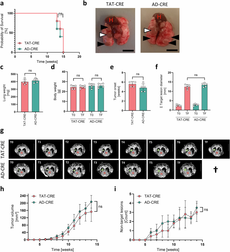
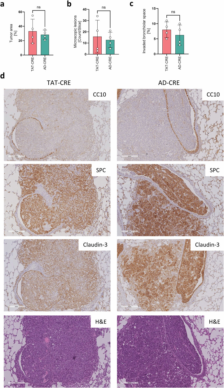
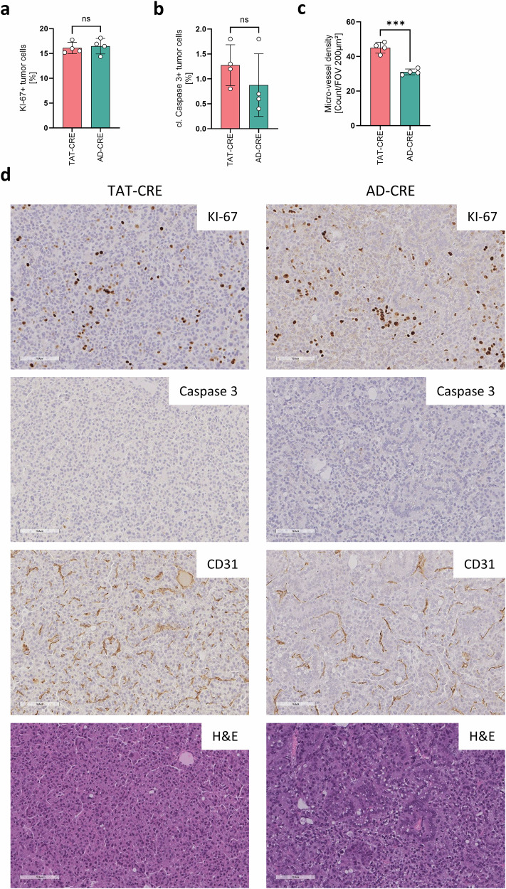
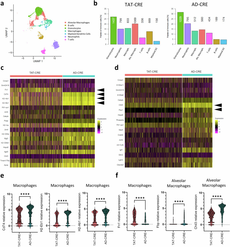
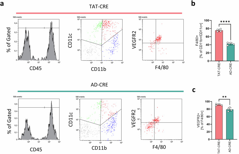
Cre-recombinase inducible model systems are extensively used in cancer research to manipulate gene expression in specific tissues and induce autochthonous tumor growth. These systems often involve the cross-breeding of genetically engineered organisms containing loxP-flanked alleles with those expressing Cre-recombinase. This approach, while effective, has the challenge of requiring high numbers of animals due to breeding requirements. Other frequently used tumor induction methods in cancer research involve the direct application of viral Cre-recombinase vectors. This approach presents the challenge of the accessibility of facilities that meet the necessary safety level. In this context, we perform a comprehensive comparison between TAT-CRE (biosafety level S1) and adenoviral Cre-recombinase induced (biosafety level S2) lung adenocarcinomas driven by KrasG12D expression and Trp53 depletion. We use in vivo lung tumor monitoring via computed tomography, single-cell RNA sequencing, immunohistochemistry and flow cytometry to elucidate similarities and differences between TAT-CRE and adenoviral Cre-recombinase induced lung adenocarcinomas. TAT-CRE induced lung tumors present differences in micro-vessels and macrophages but with corresponding tumor onset and growth characteristics compared to adenoviral-Cre recombinase induced lung tumors. Taken together, TAT-CRE is a valuable genetic engineering safety level S1 alternative for cancer induction and may be implemented in other cancer models than lung cancer.


Conflict of interest statement

Competing interests: The authors declare no competing interests.

Figures

Fig. 1
Fig. 1. Lung cancer induced by TAT-CRE and AD-CRE showed corresponding macroscopic tumor growth parameters.
Lung adenocarcinomas driven by a CRE-inducible conditional KrasG12D mutant and functional Trp53 knock out were induced by TAT-CRE (n = 5, 3 males, 2 females) and AD-CRE inhalation (n = 5, 3 males, 2 females) and macroscopically analyzed. a Probability of survival was analyzed as Kaplan-Meier-Curves and statistically evaluated using the Mantel-Cox test. b Picture of final stage lung adenocarcinomas indicating the target lesion (white), the non-target lesions (black) and the heart (H). Bars indicate 8 mm. c Lung weight was determined directly after harvest of the lungs. d Body weight at T0 (3 weeks after inhalation) and TF (final stage before lung harvest). e, f Tumor onset and target lesion diameter at T0 (3 weeks after inhalation) and TF (final stage before lung harvest) lesions were determined based on µCT DICOM data analysis using OsiriX. c-f p-values were calculated using an unpaired two-sided t-test. Data is shown as mean ± SD. P-values ≤ 0.05 are considered as significant. g Representative weekly µCT measurements starting 3 weeks after CRE inhalation. Target lesion and heart (H) are indicated. h, i Tumor volume and non-target lesions were determined based on µCT DICOM data analysis using OsiriX. Statistical analysis was done using ANOVA. P-values ≤ 0.05 are considered as significant.
Fig. 2
Fig. 2. TAT-CRE induced lung tumors expressed CC10 more frequently than AD-CRE induced lung tumors.
Lung adenocarcinomas induced by TAT-CRE (n = 4, 2 males, 2 females) and AD-CRE (n = 4, 2 males, 2 females) inhalation were analyzed for pulmonary markers by IHC. a The average staining intensity of Claudin-3 was calculated taking into account the average of all tumor lesions per slice. b Tumor lesions were classified as SPC+; CC10+/SPC+ and CC10+, respectively. A tumor lesion was defined by a minimal diameter of 100 µm. Tumor lesions are indicated in the H&E stain by a white dashed line. p-values were calculated using an unpaired two-sided t-test. Data is shown as mean ± SD. P-values ≤ 0.05 are considered as significant. c Representative IHC stain of CC10, SPC, Claudin-3 and H&E, magnification 6x. Bars indicate 400 µm.
Fig. 3
Fig. 3. Lung cancer induced by TAT-CRE and AD-CRE had a comparable capacity to invade the bronchiolar space.
Lung adenocarcinomas induced by TAT-CRE (n = 4, 2 males, 2 females) and AD-CRE (n = 4, 2 males, 2 females) inhalation were analyzed regarding bronchiolar invasion based on IHC stains. a, b Tumor area and microscopic lesions was determined based on Claudin-3 positive tumor lesions normalized to total lung area per slice. A tumor lesion was defined by a minimal diameter of 100 µm. c Invaded bronchiolar space was determined based on the evaluation of CC10 and SPC IHC stains, taking into account the average of all detectable bronchiolar spaces per slice. d Representative IHC stains of CC10, SPC, Claudin-3 and H&E, magnification 10x. Bars indicate 200 µm. p-values were calculated using an unpaired two-sided t-test. Data is shown as mean ± SD. P-values ≤ 0.05 are considered as significant.
Fig. 4
Fig. 4. TAT-CRE induced lung tumors showed significantly increased micro-vessels compared to AD-CRE induced lung tumors.
Lung adenocarcinomas induced by TAT-CRE (n = 4, 2 males, 2 females) and AD-CRE (n = 4, 2 males, 2 females) inhalation were analyzed regarding cell proliferation, micro-vessel density and apoptosis based on IHC stains. a KI-67 stain was analyzed to determine the cell proliferation rate, taking into account the average of all tumor lesions per slice. b Cleaved Caspase 3 stain was analyzed to determine the apoptosis rate, taking into account the average of all tumor lesions per slice. c CD31 stain was analyzed to determine the micro-vessel density per field of view (FOV) of 200 µm². d Representative IHC stains of KI-67, CD31, cleaved Caspase-3 and H&E, magnification 20x. Bars indicate 100 µm. p-values were calculated using an unpaired two-sided t-test. Data is shown as mean ± SD. P-values ≤ 0.05 are considered as significant.
Fig. 5
Fig. 5. TAT-CRE induced lung tumors harbored a considerable higher fraction of endothelial cells and fibroblasts than AD-CRE induced lung tumors.
a Uniform manifold approximation and projection (UMAP) representation of the integrated scRNA-seq data of TAT-CRE and AD-CRE induced lung adenocarcinoma samples (n = 1 per group, 2 males), colored by dataset (left) and by cell category (right). Each dot represents a single cell. b UMAP indicating lineage marker expression of Ptprc (immune), Epcam (epithelial), Col1a1 (fibroblasts) and Cldn5 (endothelial). c UMAP visualization of the integrated scRNA-seq data of TAT-CRE and AD-CRE induced lung adenocarcinoma samples, colored by inferred cell type. d Stacked bar plot showing the fraction per condition (TAT-CRE vs AD-CRE) among non-immune cell types: AT1 (alveolar type 1) cells, AT2 (alveolar type 2) cells, Club cells, ciliated cells, endothelial cells and fibroblasts. e UMAP representation of the integrated scRNA-seq data of epithelial cells only, colored by cell type. f Relative amount of epithelial cell types among epithelial cells identified for TAT-CRE and AD-CRE induced lung adenocarcinoma samples, respectively, with numbers indicating the corresponding absolute cell numbers. g UMAP representation of the integrated scRNA-seq data of epithelial cells only, colored by malignancy.
Fig. 6
Fig. 6. Pro-tumorigenic macrophages were more abundant in TAT-CRE vs AD-CRE induced lung tumors.
a UMAP representation of the integrated scRNA-seq data of immune cells only for TAT-CRE and AD-CRE induced lung adenocarcinoma samples (n = 1 per group, 2 males), colored by identified cell types. Each dot represents a single cell. b Relative amount of immune cell types among immune cells identified for TAT-CRE and AD-CRE induced lung adenocarcinoma samples, respectively, with numbers indicating the corresponding absolute cell numbers. c, d Heatmap showing the top 20 differentially expressed genes (DEGs) in macrophages and alveolar macrophages, respectively. Arrows indicate MHC-II-related and angiogenesis-related genes. e Differentially expressed MHC-II-related genes in macrophages of TAT-CRE and AD-CRE induced lung adenocarcinoma samples. f Differentially expressed angiogenesis-related genes in macrophages and alveolar macrophages of TAT-CRE and AD-CRE induced lung adenocarcinoma samples. p-values were calculated using an unpaired two-sided t-test. Data is shown as mean ± SD. P-values ≤ 0.05 are considered as significant.
Fig. 7
Fig. 7. TAT-CRE induced lung tumors harbored an increased fraction of VEGFR2 positive macrophages compared to AD-CRE induced lung tumors.
Lung adenocarcinomas induced by TAT-CRE (n = 4, 2 males, 2 females) and AD-CRE (n = 4, 2 males, 2 females) inhalation were analyzed by flow cytometry. a Representative histograms, dot plots and the gating of CD45, CD11c, CD11b, F4/80 and VEGFR2 stains. b F4/80 was analyzed to determine the macrophage rate in the CD11+/CD11b+ immune cell population. c VEGFR2 was analyzed as a pro-angiogenic marker on macrophages. p-values were calculated using an unpaired two sided t-test. Data is shown as mean ± SD. P-values ≤ 0.05 are considered as significant.

Similar articles

References

    1. Misawa, H. et al. Type selective ablation of postnatal slow and fast fatigue-resistant motor neurons in mice induces late onset kinetic and postural tremor following fiber-type transition and myopathy. Exp. Neurol.376, 114772 (2024). - PubMed
    1. Chen, Y. et al. Cancer-Associated Endocrine Cells Participate in Pancreatic Carcinogenesis. Gastroenterology10.1053/j.gastro.2024.07.016 (2024). - PubMed
    1. Alvarez-Martinez, M. et al. Blimp-1 and c-Maf regulate immune gene networks to protect against distinct pathways of pathobiont-induced colitis. Nat. Immunol.25, 886–901 (2024). - PMC - PubMed
    1. Sutherland, K. D. et al. Cell of origin of small cell lung cancer: inactivation of Trp53 and Rb1 in distinct cell types of adult mouse lung. Cancer Cell19, 754–764 (2011). - PubMed
    1. Kun, E. H. S. et al. Differences in gynecologic tumor development in Amhr2-Cre mice with KRAS(G12D) or KRAS(G12V) mutations. Sci. Rep.10, 20678 (2020). - PMC - PubMed