Correlation between basal cell adenoma and basal cell adenocarcinoma of the salivary gland: a histomorphological and molecular review of 129 cases
- PMID: 40360858
- PMCID: PMC12289828
- DOI: 10.1007/s00428-025-04120-7
Correlation between basal cell adenoma and basal cell adenocarcinoma of the salivary gland: a histomorphological and molecular review of 129 cases
Abstract
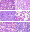
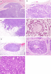
Basal cell adenoma (BCA) and basal cell adenocarcinoma (BCAC) are salivary gland tumors with biphasic differentiation, composed of luminal ductal cells and abluminal basal cells with a high nuclear-to-cytoplasmic ratio. While BCA is a relatively common benign tumor, BCAC is a rare malignancy, and its genetic context and relationship with BCA remain unclear. We investigated 93 BCA and 36 BCAC cases to further characterize these two tumor entities from histological and molecular perspectives. BCA/BCAC proliferated in a mixture of tubular, trabecular, solid, cribriform, and membranous patterns. A jigsaw puzzle pattern, peripheral palisading, S100-positive stroma, cystic change, and sclerosis were observed in approximately 50% of the cases. BCAC demonstrated the following malignant features: infiltration to surrounding tissue, tumor necrosis, and increased mitotic activity (81%, 22%, and 22%, respectively). The nuclear expression of β-catenin was frequently observed in both BCA and BCAC (89% and 60%), and CTNNB1 hotspot mutations were detected in 46% and 48% of BCA and BCAC cases, respectively. Tubular patterns of growth, jigsaw puzzle patterns, peripheral palisading, S100-positive stroma, and cystic changes were more common in β-catenin-positive BCA/BCAC than in β-catenin-negative BCA/BCAC. Among the β-catenin-negative BCA/BCAC cases, one case each harbored PLAG1 and MYB rearrangements. We concluded that β-catenin-positive BCA and BCAC share common histologic and molecular features, and BCAC is considered a malignant counterpart of BCA. β-Catenin-negative BCA/BCAC might include morphological mimickers, which can be genetically classified into other tumor types, including pleomorphic adenoma and adenoid cystic carcinoma.
Keywords: CTNNB1; Basal cell adenocarcinoma; Basal cell adenoma; Salivary gland tumor; β-catenin.
© 2025. The Author(s).
Conflict of interest statement
Declarations. Ethical approval: This study was approved by the institutional ethics review board of each participating institution (2018–0190). Consent to participate and consent for publication: The need to obtain informed consent was waived due to the retrospective nature of the analysis. Competing interests: The authors declare no competing interests.
Figures


Similar articles
-
Difference in transducin-like enhancer of split 1 protein expression between basal cell adenomas and basal cell adenocarcinomas - an immunohistochemical study.Diagn Pathol. 2018 Jul 27;13(1):48. doi: 10.1186/s13000-018-0726-8. Diagn Pathol. 2018. PMID: 30053869 Free PMC article.
-
Wnt/β-catenin signal alteration and its diagnostic utility in basal cell adenoma and histologically similar tumors of the salivary gland.Pathol Res Pract. 2018 Apr;214(4):586-592. doi: 10.1016/j.prp.2017.12.016. Epub 2018 Jan 3. Pathol Res Pract. 2018. PMID: 29496310
-
CTNNB1 mutations in basal cell adenoma of the salivary gland.J Formos Med Assoc. 2018 Oct;117(10):894-901. doi: 10.1016/j.jfma.2017.11.011. Epub 2017 Dec 7. J Formos Med Assoc. 2018. PMID: 29224720
-
Minor salivary gland basal cell adenocarcinoma: a systematic review and report of a new case.JAMA Otolaryngol Head Neck Surg. 2015 Mar;141(3):276-83. doi: 10.1001/jamaoto.2014.3344. JAMA Otolaryngol Head Neck Surg. 2015. PMID: 25555241
-
Basal Cell Adenoma and Basal Cell Adenocarcinoma.Surg Pathol Clin. 2021 Mar;14(1):25-42. doi: 10.1016/j.path.2020.09.005. Epub 2021 Jan 5. Surg Pathol Clin. 2021. PMID: 33526221 Review.
References
-
- WHO Classification of Tumours Editorial Board (2024) WHO classification of tumours, head and neck tumours, 5th edition. International Agency for Research on Cancer, Lyon
-
- Bishop JA, Thompson LDR, Wakely PE, Weinreb I (2021) Tumors of the salivary glands (AFIP atlases of tumor and non-tumor pathology). ARP Press, Arlington
-
- Robinson RA (2021) Basal cell adenoma and basal cell adenocarcinoma. Surg Pathol Clin 14:25–42. 10.1016/j.path.2020.09.005 - PubMed
-
- Nagao T, Sugano I, Ishida Y, Matsuzaki O, Konno A, Kondo Y, Nagao K (1997) Carcinoma in basal cell adenoma of the parotid gland. Pathol Res Pract 193:171–178. 10.1016/s0344-0338(97)80074-x - PubMed
MeSH terms
Substances
Grants and funding
LinkOut - more resources
Full Text Sources
Medical
Miscellaneous

