Evaluation of in vivo and in vitro binding property of a novel candidate PET tracer for CSF1R imaging and comparison with two currently-used CSF1R-PET tracers
- PMID: 40360942
- PMCID: PMC12075084
- DOI: 10.1186/s41181-025-00345-8
Evaluation of in vivo and in vitro binding property of a novel candidate PET tracer for CSF1R imaging and comparison with two currently-used CSF1R-PET tracers
Abstract
Background: Colony-stimulating factor 1 receptor (CSF1R) is a promising imaging biomarker for neuroinflammation and tumor-associated macrophages. However, existing positron emission tomography (PET) tracers for CSF1R imaging often suffer from limited specificity or sensitivity.
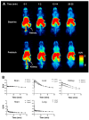
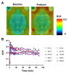
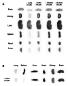
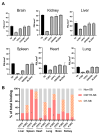
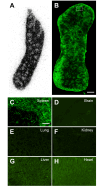
Results: We have performed 11C-labeled radiosynthesis of compound FJRD (3-((2-amino-5-(1-methyl-1H-pyrazol-4-yl)pyridin-3-yl)ethynyl)-N-(4-methoxyphenyl)-4-methylbenzamide), which exhibits excellent affinity for CSF1R, and evaluated its in vivo and in vitro binding properties. PET images of [11C]FJRD show low brain uptake and specific binding in the living organs, except the kidneys in both normal mice and rats. In vitro autoradiographs demonstrate high levels of specific binding in all investigated organs, including the brain, spleen, liver, kidneys and lungs, when self-blocking was used. The addition of CPPC partially blocked in vitro [11C]FJRD binding in these organs, with blocking effects ranging from 9 to 67%. In contrast, the other two CSF1R inhibitors, GW2580 and BLZ945, showed minimal blocking effects, suggesting unignorable off-target binding in these organs. Furthermore, specific binding of [11C]CPPC and [11C]GW2580 was faint in the mouse organs, with [11C]CPPC demonstrating detectable binding only in the spleen.
Conclusions: These results suggest that [11C]FJRD is a potential CSF1R-PET tracer for more sensitive detection of CSF1R, compared to [11C]CPPC and [11C]GW2580. However, the high level off-target binding necessitates further improvements in specificity for CSF1R imaging.
Keywords: Autoradiography; Colony-stimulating factor 1 receptor (CSF1R); Positron emission computed tomography (PET); [11C]CPPC; [11C]o-aminopyridyl alkynyl derivative.
© 2025. The Author(s).
Conflict of interest statement
Declarations. Ethics approval and consent to participate: The mice and rats in this study were maintained and handled in accordance with the National Research Council’s Guide for the Care and Use of Laboratory Animals, as well as the specific institutional guidelines. The experimental protocols involving animals were thoroughly reviewed and approved by the Animal Ethics Committees of Fudan University and the National Institutes for Quantum Science and Technology. These approvals ensure that all procedures complied with ethical standards for the humane treatment of animals, reflecting the researchers’ commitment to ethical scientific practices and the welfare of the animals involved in the study. Consent for publication: Not applicable. Competing interests: The authors declare that they have no competing interests.
Figures



Update of
-
Evaluation of in-vivo and in-vitro binding property of a novel PET tracer for CSF1R imaging and comparison with two currently-used CSF1R-PET tracers.Res Sq [Preprint]. 2025 Mar 20:rs.3.rs-6194254. doi: 10.21203/rs.3.rs-6194254/v1. Res Sq. 2025. Update in: EJNMMI Radiopharm Chem. 2025 May 13;10(1):23. doi: 10.1186/s41181-025-00345-8. PMID: 40166008 Free PMC article. Updated. Preprint.
Similar articles
-
Evaluation of in-vivo and in-vitro binding property of a novel PET tracer for CSF1R imaging and comparison with two currently-used CSF1R-PET tracers.Res Sq [Preprint]. 2025 Mar 20:rs.3.rs-6194254. doi: 10.21203/rs.3.rs-6194254/v1. Res Sq. 2025. Update in: EJNMMI Radiopharm Chem. 2025 May 13;10(1):23. doi: 10.1186/s41181-025-00345-8. PMID: 40166008 Free PMC article. Updated. Preprint.
-
PET imaging of colony-stimulating factor 1 receptor: A head-to-head comparison of a novel radioligand, 11C-GW2580, and 11C-CPPC, in mouse models of acute and chronic neuroinflammation and a rhesus monkey.J Cereb Blood Flow Metab. 2021 Sep;41(9):2410-2422. doi: 10.1177/0271678X211004146. Epub 2021 Mar 24. J Cereb Blood Flow Metab. 2021. PMID: 33757319 Free PMC article.
-
Discovery of a High-Affinity Fluoromethyl Analog of [11C]5-Cyano-N-(4-(4-methylpiperazin-1-yl)-2-(piperidin-1-yl)phenyl)furan-2-carboxamide ([11C]CPPC) and Their Comparison in Mouse and Monkey as Colony-Stimulating Factor 1 Receptor Positron Emission Tomography Radioligands.ACS Pharmacol Transl Sci. 2023 Mar 10;6(4):614-632. doi: 10.1021/acsptsci.3c00003. eCollection 2023 Apr 14. ACS Pharmacol Transl Sci. 2023. PMID: 37082755 Free PMC article.
-
Candidate Tracers for Imaging Colony-Stimulating Factor 1 Receptor in Neuroinflammation with Positron Emission Tomography: Issues and Progress.ACS Pharmacol Transl Sci. 2023 Oct 18;6(11):1632-1650. doi: 10.1021/acsptsci.3c00213. eCollection 2023 Nov 10. ACS Pharmacol Transl Sci. 2023. PMID: 37974622 Free PMC article. Review.
-
Efficiency gains in tracer identification for nuclear imaging: can in vivo LC-MS/MS evaluation of small molecules screen for successful PET tracers?ACS Chem Neurosci. 2014 Dec 17;5(12):1154-63. doi: 10.1021/cn500073j. Epub 2014 Oct 13. ACS Chem Neurosci. 2014. PMID: 25247893 Review.
References
-
- Adhikari A, Chauhan K, Adhikari M, Tiwari AK. Colony stimulating Factor-1 receptor: an emerging target for neuroinflammation PET imaging and AD therapy. Bioorg Med Chem. 2024;100:117628. - PubMed
-
- Altomonte S, Yan X, Morse CL, Liow JS, Jenkins MD, Montero Santamaria JA, Zoghbi SS, Innis RB, Pike VW. Discovery of a High-Affinity fluoromethyl analog of [(11)C]5-Cyano-N-(4-(4-methylpiperazin-1-yl)-2-(piperidin-1-yl)phenyl)furan-2-carboxamide ([(11)C]CPPC) and their comparison in mouse and monkey as Colony-Stimulating factor 1 receptor positron emission tomography radioligands. ACS Pharmacol Transl Sci. 2023;6(4):614–32. - PMC - PubMed
-
- An X, Wang J, Tong L, Zhang X, Fu H, Zhang J, Xie H, Huang Y, Jia H. (18)F-Labeled o–aminopyridyl alkynyl radioligands targeting colony-stimulating factor 1 receptor for neuroinflammation imaging. Bioorg Med Chem. 2023;83:117233. - PubMed
Grants and funding
LinkOut - more resources
Full Text Sources
Research Materials
Miscellaneous
