. 2025 Apr 23;15(9):1207.
doi: 10.3390/ani15091207.

Antiviral Activity of 1-Deoxynojirimycin Extracts of Mulberry Leaves Against Porcine Epidemic Diarrhea Virus

Affiliations

Antiviral Activity of 1-Deoxynojirimycin Extracts of Mulberry Leaves Against Porcine Epidemic Diarrhea Virus

Yiwei Sun et al. Animals (Basel). .

Abstract

Porcine epidemic diarrhea virus (PEDV), a highly infectious alphacoronavirus, has resulted in substantial economic losses within the global swine industry. Existing vaccines and therapeutic agents have proven inadequate in effectively preventing and controlling PEDV. Natural compounds offer distinct advantages in antiviral research due to their abundant availability, diverse biological activities, and low toxicity. In this study, the antiviral properties of the naturally occurring alkaloid 1-deoxynojirimycin (DNJ) against PEDV were examined. The CC50 of DNJ was determined to be 912.5 μM through experimental analysis on Vero-E6 cells. DNJ demonstrated an inhibitory effect on PEDV activity, with a 50% inhibitory concentration (IC50) of 57.76 μM. The compound primarily inhibited PEDV proliferation during the viral life cycle stages of attachment and replication. Moreover, DNJ mitigated the production of reactive oxygen species (ROS) and inflammation associated with PEDV infection. Computational docking predictions suggest that the viral non-structural proteins include Nsp12, Nsp14, and Nsp16 may serve as potential targets for DNJ. Consequently, DNJ represents a promising candidate for the development of novel therapeutic agents against PEDV.

Keywords: 1-deoxynojirimycin (DNJ); PEDV; antiviral activity.


Conflict of interest statement

The authors declare that they have no conflict of interest.

Figures

Figure 1
Figure 1
The chemical structure, CCK8, and IC50 of DNJ. (A) The chemical structure of DNJ. (B) Morphological changes were observed after the cells were treated with DNJ at different concentrations for 12 h using a fluorescence microscope under bright-field. (C) Vero E6 cells were treated with 0 μM, 50 μM, 100 μM, 200 μM, 400 μM, 600 μM, and 800 μM DNJ for 12 h, and then treated with CCK 8 for 2 h. The OD450 value was measured, and nonlinear regression analysis was performed to calculate the CC50 value. (D) The half maximal inhibitory concentration (IC50) was determined by cell ELISA, and the IC50 was calculated by nonlinear regression analysis.
Figure 2
Figure 2
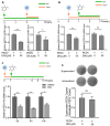
DNJ inhibits PEDV infection in Vero-E6 cells. (A) The overall design of Vero E6 cells infected and treated with DNJ (25 μM, 50 μM, 100 μM). The green line indicates drug treatment, and the orange line indicates PEDV infection. (B) Samples were collected at 6, 12, and 24 h post-infection with SQ2014 (MOI = 0.1), and the protein levels of PEDV-N and actin were detected by Western blot. (C) After 12 h of infection, the protein samples with different concentrations of DNJ were detected by Western blot. (D) Quantitative detection of PEDV S and N mRNA level by qRT-PCR. (E) The cells were collected 12 h after infection, and the virus titer of SQ2014 was detected by PFU. The means and standard deviations of three independent experimental replicates are shown. ** p < 0.01; *** p < 0.001.
Figure 3
Figure 3
The inhibition stage of DNJ on PEDV. (A) This section describes the comprehensive approach for infecting Vero-E6 cells and administering DNJ at a concentration of 100 µM. The treatment procedure is divided into three separate phases: pre-treatment, co-treatment, and post-treatment. (B) In accordance with the treatment protocol depicted in (A), Western blot analysis was conducted to evaluate the protein levels of both the virus and actin. (C) DNJ directly targets PEDV SQ2014 experimental results.
Figure 4
Figure 4
Effects of DNJ on the attachment, entry, replication, and release of PEDV. (A) The experimental design of DNJ and PEDV infection cycle stage treatment of Vero E6 cells. The green line segment indicates drug treatment, and the orange line segment indicates PEDV infection. Virus attachment experiment: the mRNA levels of PEDV S and N were detected by qRT-PCR after 1 h of infection with PEDV in DNJ-treated Vero E6 cells. (B) Virus entry experiment: PEDV S and N mRNA levels were detected by qRT-PCR 2 h after PEDV infection in Vero E6 cells treated with DNJ. (C) Virus replication experiment: Vero E6 cells were treated with DNJ within 6, 8, and 10 h post-infection, and the levels of PEDV N mRNA were detected by qRT-PCR. (D) Virus release assay: cells and supernatants were collected after virus infection, and PEDV titers in cells and supernatants were detected by PFU. The means and standard deviations of three independent experimental replicates are shown. * p < 0.05; ** p < 0.01; *** p < 0.001; ns, not significant.
Figure 5
Figure 5
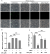
DNJ alleviates PEDV by inhibiting ROS production. (A) Vero cells were infected with PEDV (MOI = 0.1). After sufficient washing, the culture medium was replaced with different concentrations of DNJ to continue culturing. After 24 h, DCFH-DA was added and incubated for 40 min to detect cell ROS. (B) Production of MDA. (C) GSH-Px activity. The means and standard deviations of three independent experimental replicates are shown. ** p < 0.01; *** p < 0.001; ns, not significant. DNJ: 1-deoxynojirimycin (DNJ); PEDV: porcine epidemic diarrhea virus; ROS: reactive oxygen species.
Figure 6
Figure 6
DNJ inhibited the expression of inflammatory cytokines and antioxidant indexes induced by PEDV infection. Vero E6 cells were infected with PEDV (MOI = 0.1) for 12 h with DNJ treatment. (A) The mRNA levels of IL-1β, (B) IL-8, (C) MCP, and (D) TNF-α were detected by RT-qPCR. The means and standard deviations of three independent experimental replicates are shown. *** p < 0.001; ns, not significant.
Figure 7
Figure 7
Molecular docking predicted the target of DNJ. The docking conformation of DNJ with (A) PEDV 3C Like Protease, (B) PEDV papain-like protease 2, (C) PEDV-S1, (D) PEDV Nsp12, (E) PEDV Nsp13, (F) PEDV Nsp14, (G) PEDV Nsp15, and (H) PEDV Nsp16. In this representation, the left diagram of each protein molecular complex is a 3D diagram of the structural conformation of the molecule and the protein, and the right diagram is a local interaction diagram of the hydrogen bond between the molecule and the protein. The green rod-like structure is a small molecule, and the orange rod-like structure is an amino acid residue that forms a hydrogen bond interaction in the protein. The amino acid name and site information are marked on it. The yellow dotted line represents the hydrogen bond, and the number next to the dotted line is the bond length. (I) The binding energy of the DNJ–protein complex, which was calculated using Autodock vina 1.1.2 software.

Similar articles

Cited by

References

    1. Li W., Li H., Liu Y., Pan Y., Deng F., Song Y., Tang X., He Q. New variants of porcine epidemic diarrhea virus, China, 2011. Emerg. Infect. Dis. 2012;18:1350–1353. doi: 10.3201/eid1803.120002. - DOI - PMC - PubMed
    1. Zhang H., Zou C., Peng O., Ashraf U., Xu Q., Gong L., Fan B., Zhang Y., Xu Z., Xue C., et al. Global Dynamics of Porcine Enteric Coronavirus PEDV Epidemiology, Evolution, and Transmission. Mol. Biol. Evol. 2023;40:msad052. doi: 10.1093/molbev/msad052. - DOI - PMC - PubMed
    1. Lin F., Zhang H., Li L., Yang Y., Zou X., Chen J., Tang X. PEDV: Insights and Advances into Types, Function, Structure, and Receptor Recognition. Viruses. 2022;14:1744. doi: 10.3390/v14081744. - DOI - PMC - PubMed
    1. Liu C., Kong N., Liu H., Zhang Y., Qin W., Zhao W., Yang X., Wang Y., Cao X., Liu T., et al. FSTL1 and TLR4 interact with PEDV structural proteins to promote virus adsorption to host cells. J. Virol. 2025;99:e0183724. doi: 10.1128/jvi.01837-24. - DOI - PMC - PubMed
    1. Wei X., She G., Wu T., Xue C., Cao Y. PEDV enters cells through clathrin-, caveolae-, and lipid raft-mediated endocytosis and traffics via the endo-/lysosome pathway. Vet. Res. 2020;51:10. doi: 10.1186/s13567-020-0739-7. - DOI - PMC - PubMed

LinkOut - more resources