. 2025 Apr 22;26(9):3963.
doi: 10.3390/ijms26093963.

Resveratrol's Pro-Apoptotic Effects in Cancer Are Mediated Through the Interaction and Oligomerization of the Mitochondrial VDAC1

Affiliations

Resveratrol's Pro-Apoptotic Effects in Cancer Are Mediated Through the Interaction and Oligomerization of the Mitochondrial VDAC1

Tal Raviv et al. Int J Mol Sci. .

Abstract

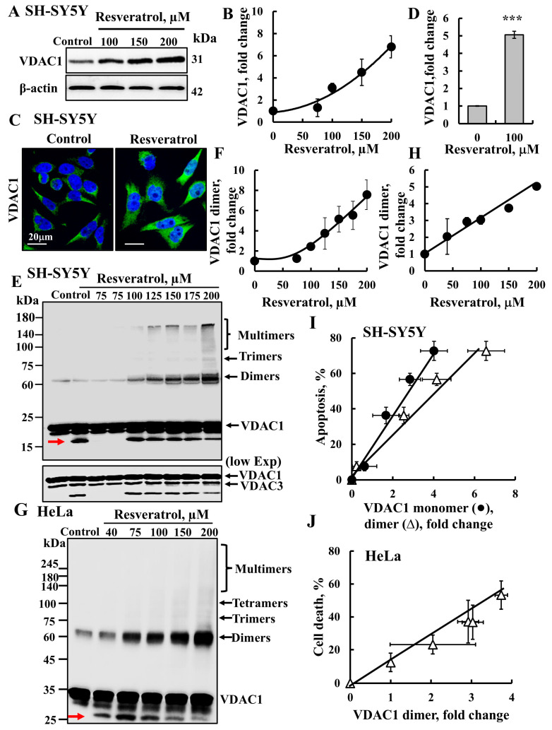
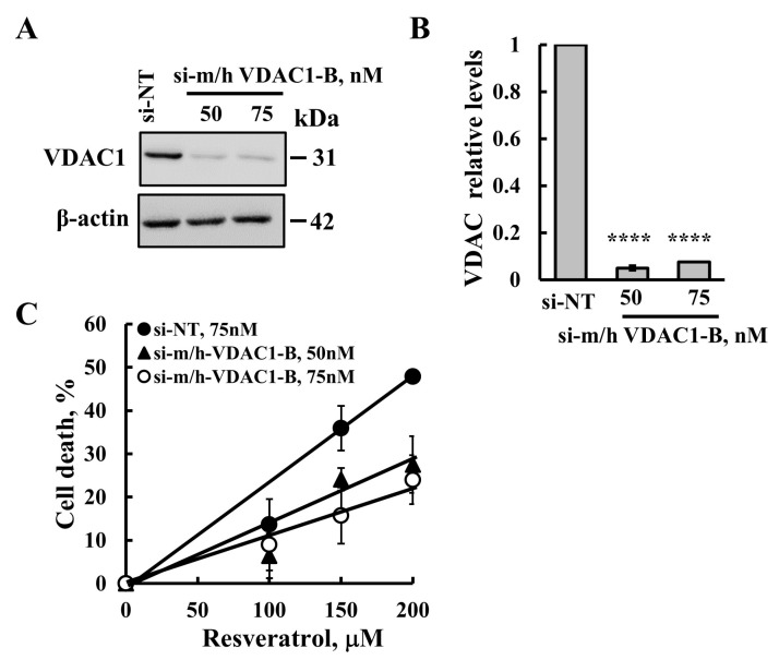
Resveratrol is a naturally occurring phenolic compound found in various foods such as red wine, chocolate, peanuts, and blueberries. Both in-vitro and in-vivo studies have shown that it has a broad spectrum of pharmacological effects such as providing cellular protection and promoting longevity. These effects include antioxidant, anti-inflammatory, neuroprotective, and anti-viral properties, as well as improvements in cardio-metabolic health and anti-aging benefits. Additionally, resveratrol has demonstrated the ability to induce cell death and inhibit tumor growth across different types and stages of cancer. However, the dual effects of resveratrol-acting to support cell survival in some contexts, while inducing cell death in others-is still not fully understood. In this study, we identify a novel target for resveratrol: the voltage-dependent anion channel 1 (VDAC1), a multi-functional outer mitochondrial membrane protein that plays a key role in regulating both cell survival and death. Our findings show that resveratrol increased VDAC1 expression levels and promoted its oligomerization, leading to apoptotic cell death. Additionally, resveratrol elevated intracellular Ca2+ levels and enhanced the production of reactive oxygen species (ROS). Resveratrol also induced the detachment of hexokinase I from VDAC1, a key enzyme in metabolism, and regulating apoptosis. When VDAC1 expression was silenced using specific siRNA, resveratrol-induced cell death was significantly reduced, indicating that VDAC1 is essential for its pro-apoptotic effects. Additionally, both resveratrol and its analog, trans-2,3,5,4'-tetrahydroxystilbene-2-O-glucoside (TSG), directly interacted with purified VDAC1, as revealed by microscale thermophoresis, with similar binding affinities. However, unlike resveratrol, TSG did not induce VDAC1 overexpression or apoptosis. These results demonstrate that resveratrol-induced apoptosis is linked to increased VDAC1 expression and its oligomerization. This positions resveratrol not only as a protective agent, but also as a pro-apoptotic compound. Consequently, resveratrol offers a promising therapeutic approach for cancer, with potentially fewer side effects compared to conventional treatments, due to its natural origins in plants and food products.

Keywords: VDAC1; apoptosis; hexokinase; mitochondria; resveratrol.


Conflict of interest statement

The authors declare that they have no conflicts of interest.

Figures

Figure 6
Figure 6
Resveratrol and TSG directly bind to VDAC1, but only resveratrol induced apoptosis. Chemical structures of resveratrol (A) and its analog trans-2,3,5,4′-tetrahydroxystilbene-2-O-glucoside (TSG) (B). SH-SY5Y cells were incubated for 24 h with the indicated concentrations of resveratrol or TSG and then analyzed for cell death using PI staining and flow cytometry (C), and for cell viability, XTT was used (D). Cells were also analyzed for VDAC1 expression levels (E) by immunoblotting using anti-VDAC1-specific antibodies. Immunoblotting with β-actin as a loading control is also shown. The levels of VDAC1 were quantified (F). In addition, cells were analyzed for VDAC1 oligomerization by incubation (1 mg/mL protein) with the cross-linking reagent EGS (100 μM; 15 min), followed by immunoblotting using anti-VDAC1 antibodies. The positions of the VDAC1 monomers and dimers are indicated (G) the red arrow points to monomeric VDAC1 undergoing intramolecular cross-linking. The level of the VDAC1 dimers was analyzed using ImageJ software (version 1.54p) (H) and is presented relative to its levels in control cells. Results represent the means ± SEM (n = 3); p < 0.05 (*), p < 0.01 (**).
Figure 1
Figure 1
Resveratrol induces apoptosis and reduces the viability of cancer cells. SH-SY5Y, HeLa, or HEK-293 cells were treated with the indicated concentrations of resveratrol for 24 h or 48 h. Then, cells were harvested and subjected to an apoptotic cell death analysis (AD) or cell viability analysis (E). (A,B) Apoptosis of SH-SY5Y and HEK-293 cells was assayed using annexin V–FITC and PI staining, and flow cytometry and cell death of HeLa cells (C) was assayed with PI staining and flow cytometry. Representative histograms of resveratrol induction of apoptosis in SH-SY5Y cells are shown (A). Quantitative analyses of three independent experiments as in A are shown (B,C). Summary of the IC50 and maximal cell death obtained for the three cell types (D). Cell viability of the SH-SY5Y cells was determined using an XTT-based assay, and viable cells were measured at 490 nm in a microplate reader (E). The results are the mean ± SEM (n = 3).
Figure 2
Figure 2
Resveratrol enhances VDAC1 expression levels and oligomerization. (A,B) SH-SY5Y cells were incubated for 24 h with the indicated concentrations of resveratrol and then analyzed for VDAC1 expression level by immunoblotting using anti-VDAC1 antibodies (A). Immunoblotting with β-actin as a loading control is also shown. Quantification of VDAC1 levels (B). (C) SH-SY5Y were seeded on 13-mm glass coverslips, treated with resveratrol (100 μM, 24 h), fixed and subjected to immunofluorescence (IF) using anti-VDAC1 antibodies. (D) Quantification of VDAC1 levels of the IF staining. (E,F) SH-SY5Y cells were incubated for 24 h with the indicated concentrations of resveratrol and analyzed for VDAC1 oligomerization by incubation (3 mg/mL protein) with the cross-linking reagent EGS (300 μM), followed by immunoblotting with anti-VDAC1 antibodies. The positions of the VDAC1 monomers, dimers, trimers, tetramers, and higher oligomers are indicated. The red arrow points to monomeric VDAC1 undergoing intramolecular cross-linking. The level of VDAC1 dimers was analyzed using ImageJ software (version 1.54v, Bethesda, MD, USA) and is presented relative to its levels in the control cells (F). (G,H) HeLa cells were incubated for 48 h with the indicated concentrations of resveratrol and analyzed for VDAC1 oligomerization (1 mg/mL protein), and incubated with EGS (100 μM), followed by immunoblotting (G) and dimer quantification (H). (I,J) Correlation plot of apoptosis percentage relative to the increase in VDAC1 monomers (●) and its dimer (Δ) levels obtained at the same concentration of resveratrol for SH-SY5Y (I) or HeLa (J). Results represent the means ± SEM; p < 0.001 (***).
Figure 3
Figure 3
Resveratrol elevates cellular Ca2+ levels and increases ROS production. SH-SY5Y cells were incubated for 24 h with the indicated concentrations of resveratrol. They were then harvested, and intracellular calcium ([Ca2+]i) levels (A,B), mitochondrial, and cellular superoxide (ROS) levels (CE) were measured using Fluo-4-AM, MitoSOX Red, and DCF, respectively, and analyzed by flow cytometry. Representative FACS histograms (A,C) and quantification (B,D,E) are presented. (F) Cells were treated as in A, and then analyzed for VDAC1 or SIRT1 expression levels by immunoblotting using anti-VDAC1 or anti-SIRT1 specific antibodies. Immunoblotting with β-actin as a loading control is also shown. (G) Quantification of VDAC1 and SIRT1 levels. Results represent the means ± SEM (n = 3); p < 0.05 (*), p < 0.01 (**), p < 0.001 (***).
Figure 4
Figure 4
Resveratrol induces HK-I detachment from the mitochondria. SH-SY5Y cells were seeded on 13-mm glass coverslips and then incubated for 24 h with resveratrol (100 μM) followed by IF staining with anti-HK-I antibodies. Images were visualized by confocal microscopy (A). Enlargements (a, b) of dashed squares are presented. The arrows point to cytosolic, diffused staining of HK-I. (B) Quantification of HK-I expression levels. (C) SH-SY5Y cells were treated with the indicated concentrations of resveratrol, which were then harvested and incubated on ice for 10 min with 0.005% digitonin. Following centrifugation (10,000× g, 5 min), the supernatants (cytosol) were subjected to SDS-PAGE and immunoblotting using anti-HK-I antibodies. Relative HK-I levels released to the cytosol are presented below the immunoblots relative to the control in relative units (RUs). Results represent the means ± SEM (n = 3); p < 0.001 (***).
Figure 5
Figure 5
VDAC1 silencing reduced resveratrol-induced apoptosis. SH-SY5Y cells were transfected with si-NT (75 nM) or with si-m/h-VDAC1-B (50 nM or 75 nM), using JetPrime transfection reagent, as described in the Section 4. At 24 h post transfection, cells were treated with the indicated concentrations of resveratrol for 24 h and subjected to immunoblotting for VDAC1 expression levels using anti-VDAC1 specific antibodies (A). (B) VDAC1 expression levels were quantified and are presented as relative units (RUs). (C) Cell death induced by resveratrol in SH-SY5Y treated with si-NT (●) or treated with si-m/h-VDAC1-B 50 nM (▲) or 75 nM (○), determined using PI staining and a flow cytometer analysis. Results reflect the mean ± SEM (n = 3), p ≤ 0.0001 (****). 2.5. Resveratrol and its Analog Directly Bind to VDAC1, but Only Resveratrol Induces Apoptosis.
Figure 7
Figure 7
Resveratrol and TSG directly bind to VDAC1. (A) Coomassie bluestained purified VDAC1 was fluorescently labeled using a NanoTemper Protein-Labeling Kit BLUE. Labeled VDAC1 was incubated for 30 min with the indicated concentrations of resveratrol, and then 3–5 μL of the samples were loaded into MSTgrade glass capillaries, and the thermophoresis process was measured. (B) Schematic presentation of the MST assay. Thermal-migration curves of VDAC1 in the presence of different concentrations of resveratrol upon starting heat (On) and stopping heating (Off). (C) VDAC1 binding in the presence of resveratrol (●) or TGS (Δ) derived from the MST measurements are presented. Results represent the means ± SEM (n = 3).
Figure 8
Figure 8
Proposed model for resveratrol inducing VDAC1 overexpression, oligomerization, and apoptosis. Resveratrol entering the cell enhances VDAC1 expression levels with the overexpressed VDAC1, shifting the equilibrium to the VDAC1 oligomeric state. This mediates the release of Ca2+, ROS, and apoptogenic proteins, leading to apoptosis. Resveratrol detaches HK-I from VDAC1, further enhancing VDAC1 oligomerization, which results in apoptosis and disrupted metabolism.

References

    1. Huminiecki L., Horbanczuk J. The functional genomic studies of resveratrol in respect to its anti-cancer effects. Biotechnol. Adv. 2018;36:1699–1708. doi: 10.1016/j.biotechadv.2018.02.011. - DOI - PubMed
    1. Pezzuto J.M. Resveratrol: Twenty Years of Growth, Development and Controversy. Biomol. Ther. 2019;27:1–14. doi: 10.4062/biomolther.2018.176. - DOI - PMC - PubMed
    1. Ramirez-Garza S.L., Laveriano-Santos E.P., Marhuenda-Munoz M., Storniolo C.E., Tresserra-Rimbau A., Vallverdu-Queralt A., Lamuela-Raventos R.M. Health Effects of Resveratrol: Results from Human Intervention Trials. Nutrients. 2018;10:1892. doi: 10.3390/nu10121892. - DOI - PMC - PubMed
    1. Dybkowska E., Sadowska A., Swiderski F., Rakowska R., Wysocka K. The occurrence of resveratrol in foodstuffs and its potential for supporting cancer prevention and treatment. A Rev. Rocz. Panstw. Zakl. Hig. 2018;69:5–14. - PubMed
    1. Wang F., Chatterjee S. Dominant Carbons in trans- and cis-Resveratrol Isomerization. J. Phys. Chem. B. 2017;121:4745–4755. doi: 10.1021/acs.jpcb.7b02115. - DOI - PubMed

MeSH terms

LinkOut - more resources