Review
. 2025 Apr 23;26(9):3998.
doi: 10.3390/ijms26093998.

Probing Peptide Assembly and Interaction via High-Resolution Imaging Techniques: A Mini Review

Affiliations
Review

Probing Peptide Assembly and Interaction via High-Resolution Imaging Techniques: A Mini Review

Xiaoming Zhang et al. Int J Mol Sci. .

Abstract

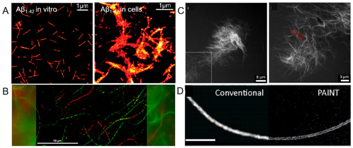
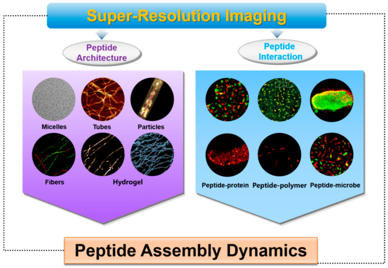
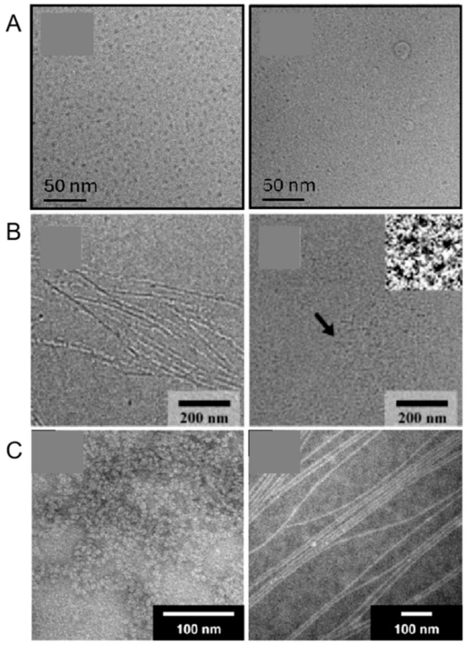
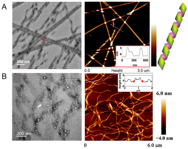
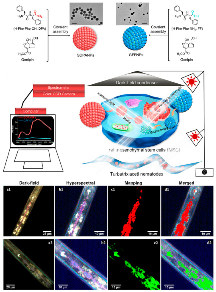
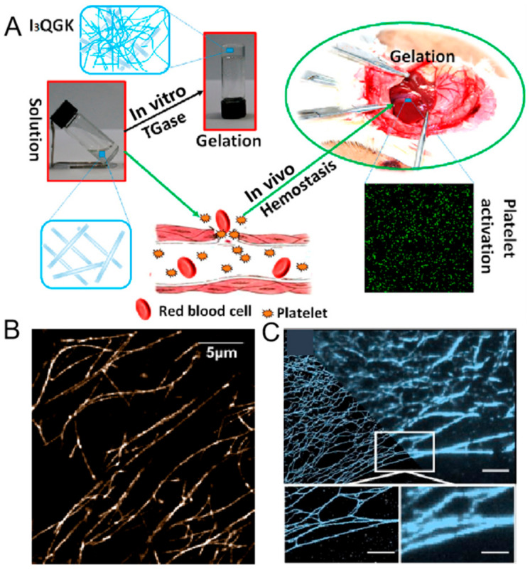
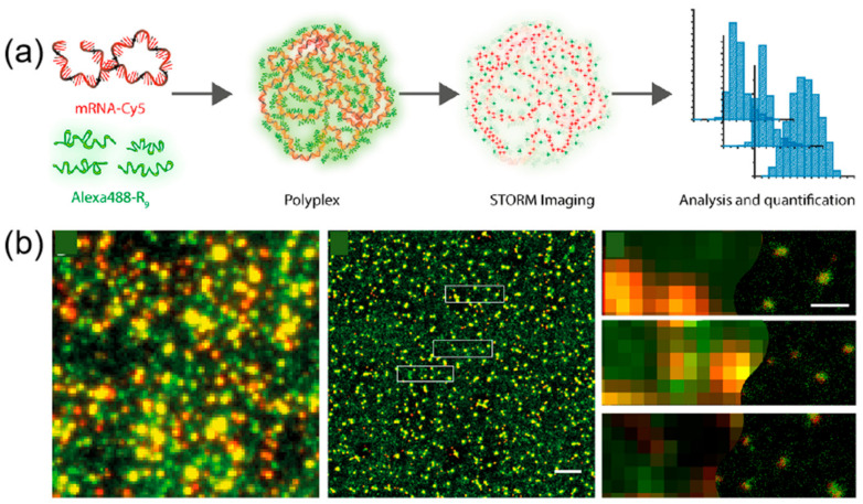
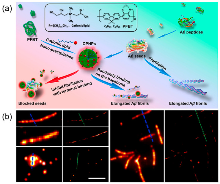
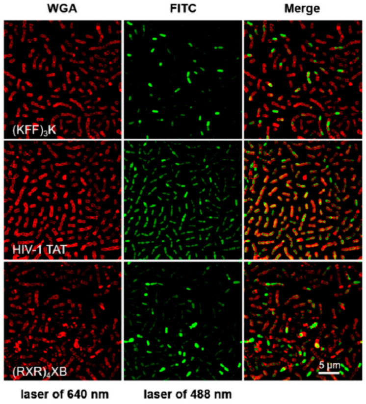
Peptide molecules, as fundamental structural units in biological systems, play pivotal roles in diverse biological processes and have garnered substantial attention in biomolecular self-assembly research. Their structural simplicity and high design flexibility make peptides key players in the development of novel biomaterials. High-resolution imaging techniques have provided profound insights into peptide assembly. Recently, the development of cutting-edge technologies, such as super-resolution microscopy (SRM) with unparalleled spatiotemporal resolution, has further advanced peptide assembly research. These advancements enable both the mechanistic exploration of peptide assembly pathways and the rational design of peptide-based functional materials. In this mini review, we systematically examine the structural diversity of peptide assemblies, including micelles, tubes, particles, fibers and hydrogel, as investigated by various high-resolution imaging techniques, with a focus on their assembly characterization and dynamic process. We also summarize the interaction networks of peptide assemblies with proteins, polymers and microbes, providing further insight into the interactions between peptide assemblies and other molecules. Furthermore, we emphasize the transformative role of high-resolution imaging techniques in addressing long-standing challenges in peptide nanotechnology. We anticipate that this review will accelerate the advancement of peptide assembly characterization, thereby fostering the creation of next-generation functional biomaterials.

Keywords: biomaterials; nanotechnology; peptide molecules; self-assembly; super-resolution microscopy.


Conflict of interest statement

The authors declare no conflict of interest.

Figures

Figure 5
Figure 5
(A) dSTORM images of Aβ1-42 in vitro and in cells. Reprinted with permission from Ref. [62]. 2011, American Chemical Society. (B) Two-color STROM image of I3K peptide fibrils. Red represents Alexa Fluor 647 dye-labeled I3K peptide fibers, while green represents Cy3B dye-labeled I3K peptide fibers. Most fibers are either pure red or pure green, with minimal entanglement or in contact, suggesting the high stability of the I3K fibers structure and its resistance to monomer exchange. Reprinted with permission from Ref. [63]. 2017, American Chemical Society. (C) STED micrographs of self-assembled short peptide sequence FFALGLAGKK. The image on the right is a magnification of the box. Reprinted with permission from Ref. [66]. 2020, American Chemical Society. (D) Comparison of conventional TIRF imaging and PAINT imaging of an FF nanofiber. Scale bar 5 µm. Reprinted with permission from Ref. [67]. 2017, The Royal Society of Chemistry.
Figure 1
Figure 1
Schematic illustration of super-resolution imaging technology application in peptide assembly dynamics.
Figure 2
Figure 2
(A) Cryo-TEM images from 0.5 wt% aqueous solutions of lipopeptides C16-CSK4RGDS and C16-CSK4GRDS. Reprinted with permission from Ref. [56]. 2024, American Chemical Society. (B) Cryo-TEM images of peptide amphiphile (PA) nanofibers and micelles with an N-methylated glycine near the core. Arrow indicates the location of insets. Reprinted with permission from Ref. [57]. 2006, Paramonov, American Chemical Society. (C) The PA micelles formed fibers, and the solution turned into a self-supporting gel after irradiation in the presence of charge-screening calcium chloride salts. Reprinted with permission from Ref. [58]. 2008, European Peptide Society and John Wiley & Sons, Ltd.
Figure 3
Figure 3
(A) Helical intermediates of Ac-KI3VK-NH2. Red arrow indicates the folded edges of helical ribbons. Bottom right inset is the cross-section profiles of helical ribbons. Reprinted with permission from Ref. [59]. 2018, WILEY-VCH Verlag GmbH & Co. KGaA, Weinheim. (B) Self-assembled nanostructures formed by KI4K in a mixture of acetonitrile and water with a volume ratio of 20%. The insert box indicates that helical bands, with heights of approximately 2.1 nm and widths mostly ranging from 30−90 nm, are dominant in this sample. Reprinted with permission from Ref. [60]. 2015, American Chemical Society.
Figure 4
Figure 4
Self-assembled peptide nanoparticles for enhanced dark-field and hyperspectral imaging. (a1) Dark-field images in the intestines of T. aceti worms; (b1) hyperspectral images; (c1) corresponding maps; (d1) dark-field results of GDPANPs. (a2) Dark-field images in the intestines of T. aceti worms; (b2) hyperspectral images; (c2) corresponding maps; (d2) dark-field results of GFFNPs. The bright spots in the dark field plot indicate strong scattering signals from the particles. The colors in the hyperspectral plot represent light scattering intensity at different wavelengths. The mapping shows the degree and location of the match with the standard nanoparticle spectrum, using different colors to differentiate material types. Brighter colors indicate a higher degree of match with the standard nanoparticle scattering spectrum. Reprinted with permission from Ref. [61]. 2021, Elsevier B.V.
Figure 6
Figure 6
(A) Self-assembly schematic of I3QGK in clinical hemostasis. Reprinted with permission from Ref. [21]. 2016, American Chemical Society. (B) STORM image of the fully labeled I3K network, showing regions of densely cross-linked fibrils. Reprinted with permission from Ref. [68]. 2018, American Chemical Society. (C) Probe reversibly binds to fibers to form PAINT image (left) vs. low-resolution image (right). The white box indicates the zoomed-in area displayed in the subfigure. Scale bars: top image, 5 µm; bottom images, 2.5 µm. Reprinted with permission from Ref. [69]. 2020, Chemistry Europe.
Figure 7
Figure 7
(A) Super-resolution SERS imaging of AuNS-RGDFC in cancer cells. RGDFC functionalized AuNS were used as label-free SERS probes and super-resolution SERS results for fixed cells incubated with AuNS-RGDFC recorded at 10 Hz. The scale bar is 2 μm for the main image and 150 nm for the enlarged view. Reprinted with permission from Ref. [70]. 2020, American Chemical Society; (B) Schematic illustration of EpCAM assembly pattern on cell membrane. Dual-color dSTORM images revealing the spatial relationship between EpCAM (green) and CD9 (red) on MCF-7 cell membranes, with magnified views of two regions (i) and (ii) showing the co-localization of CD9 and EpCAM. The microsphere, circled in the upper left corner, was used to correct x-y drift and optical registration. Scale bars: 5 μm in main image, and 1 μm in (i−ii). Reprinted with permission from Ref. [71]. 2019, American Chemical Society.
Figure 8
Figure 8
(a) Diagram of hypothesized function of CPP-SpyCatcher fusion proteins; (b) Design of the CPP-SpyCatcher fusion; (c) Lattice SIM imaging of stained bacteria shows internalization of SpyCatcher and CPP fusions. The overlap of signals from FM4-64 dye-labeled cell membranes (red) and AF647-labeled SpyCatcher003 (yellow) indicates that the probe was successfully transduced into the cytoplasmic lysate of freeze-thawed E. coli, even in the absence of CPP sequences. Scale bars: 5 μm. Reprinted with permission from Ref. [72]. 2023, IOP Publishing Ltd.
Figure 9
Figure 9
(a) Schematic representation of polyplex formation from mRNA-Cy5 and AlexaFluor488-R9; (b) Conventional fluorescent image (red represents mRNA molecules and green R9 molecules) and dSTORM image of polyplexes. The yellow spots indicate co-localization of mRNA polyplexes with the R9 peptide. The boxed area in the middle image is magnified in the right panel. Scale bar: 2 μm (middle), 400 nm (right). Reprinted with permission from Ref. [75]. 2019, American Chemical Society.
Figure 10
Figure 10
(a) Schematic diagram of procedures for CPNPs fabrication and Aβ1−40 fibril inhibition with CPNPs; (b) Representative conventional fluorescence microscopy images of Aβ1−40 fiber filaments and corresponding reconstructed super-resolution images. Scale bar 5 μm. Reprinted with permission from Ref. [76]. 2019, American Chemical Society.
Figure 11
Figure 11
(A) AMP-2HBT for bacterial imaging and killing. The white box indicates the area enlarged in the subgraph below. Reprinted with permission from Ref. [77]. 2018, American Chemical Society. (B) Demonstration of the antimicrobial effect of LL-37-TAMRA on intracellular Mycobacterium tuberculosis using two-color STED microscopy. The white box indicates the area enlarged in the subgraph with LAM and LL-37, respectively. Reprinted with permission from Ref. [78]. 2020, MDPI.
Figure 12
Figure 12
Representative images were used for analyzing the internalization efficiency of FITC-labeled CPPs in S. suis by SR-SIM. Reprinted with permission from Ref. [79]. 2024, MDPI.

Similar articles

References

    1. Chang R., Yuan C., Zhou P., Xing R., Yan X. Peptide self-assembly: From ordered to disordered. Acc. Chem. Res. 2024;57:289–301. doi: 10.1021/acs.accounts.3c00592. - DOI - PubMed
    1. Levin A., Hakala T.A., Schnaider L., Bernardes G.J.L., Gazit E., Knowles T.P.J. Biomimetic peptide self-assembly for functional materials. Nat. Rev. Chem. 2020;4:615–634. doi: 10.1038/s41570-020-0215-y. - DOI - PMC - PubMed
    1. Lou S., Wang X., Yu Z., Shi L. Peptide tectonics: Encoded structural complementarity dictates programmable Self-Assembly. Adv. Sci. 2019;6:1802043. doi: 10.1002/advs.201802043. - DOI - PMC - PubMed
    1. Wang C., Fu L., Hu Z., Zhong Y. A mini-review on peptide-based self-assemblies and their biological applications. Nanotechnology. 2021;33:062004. doi: 10.1088/1361-6528/ac2fe3. - DOI - PubMed
    1. Hamley I.W. Small bioactive peptides for biomaterials design and therapeutics. Chem. Rev. 2017;117:14015–14041. doi: 10.1021/acs.chemrev.7b00522. - DOI - PubMed

LinkOut - more resources