Randomized Controlled Trial
. 2025 Apr 23;26(9):4001.
doi: 10.3390/ijms26094001.

Skin Photoprotection and Anti-Aging Benefits of a Combination of Rosemary and Grapefruit Extracts: Evidence from In Vitro Models and Human Study

Affiliations
Randomized Controlled Trial

Skin Photoprotection and Anti-Aging Benefits of a Combination of Rosemary and Grapefruit Extracts: Evidence from In Vitro Models and Human Study

Pau Navarro et al. Int J Mol Sci. .

Abstract

Skin exposure to ultraviolet radiation (UVR) causes oxidative stress, inflammation, and collagen degradation and can trigger erythema. While topical formulas protect the skin from UV damage, there is growing evidence that certain botanical ingredients taken orally may have an added benefit. This study evaluated the photoprotective, anti-photoaging, and anti-erythema efficacy of a combination of rosemary and grapefruit extract (Nutroxsun®). Radical oxygen species (ROS) generation and interleukin production were determined in UV-irradiated keratinocytes (HaCaT). Also, collagen and elastin secretion and metalloproteinase (MMP-1 and MMP-3) content were assessed in UV-irradiated fibroblasts (NHDFs). Furthermore, a placebo-controlled, randomized, crossover study was conducted in 20 subjects (phototypes I to III) receiving two doses, 100 and 200 mg, of the ingredient. Skin redness (a* value, CIELab) after exposure to one minimal erythemal dose of UVR was assessed. As a result, the botanical blend significantly attenuated the UVR-induced reductions of procollagen I and elastin and lowered MMP-1 and MMP-3 protein secretion. Also, a reduction in ROS and proinflammatory interleukins (IL-1, IL-8, and IL-6) was observed. Finally, the botanical blend, at both doses, significantly reduced UV-induced erythema reaction from the first day of intake and accelerated recovery. These findings reinforce the potential of this ingredient as an effective dietary solution to protect the skin against UV-induced damage.

Keywords: Citrus paradisi; Rosmarinus officinalis; collagen; dietary supplement; elastin; erythema; metalloproteinases and clinical study; photoaging; photoprotection.


Conflict of interest statement

N.C., J.J., A.G. and P.N. belong to the Research and Development Department at Monteloeder S.L. This does not alter the author’s adherence to all the journal policies on sharing data and materials.

Figures

Figure 1
Figure 1
Inhibition profile of Nutroxsun® on matrix metalloproteinases (MMPs). The inhibitory effect was measured using a fluorometric assay, and the results are expressed as the percentage inhibition of enzymatic activity. Each bar represents the mean ± SD of two experiments. IC50 values, obtained with the ATT Bioquest online tool, are indicated for each MMP.
Figure 2
Figure 2
Bar graph representing MMP-1 (a) and MMP-3 (b) content in NHDF culture supernatants, normalized to cell viability in the presence of Nutroxsun® (Nx) and upon exposure to UVR, as indicated in Section 4. The graphs are plotted as the percentage of change in MMP content compared to the non-irradiated untreated control at 100%. Data are expressed as mean ± SD. ** (p < 0.01) and *** (p < 0.001) indicate statistically significant differences compared to the non-irradiated untreated control (gray bars) and # (p < 0.05), ## (p < 0.01) and ### (p < 0.001) indicate statistically significant differences compared to the UVR-untreated controls (black bars).
Figure 3
Figure 3
Bar graph representing elastin (a) and pro-collagen I (b) content in NHDF culture supernatants, normalized to cell viability in the presence of Nutroxsun® (Nx) and upon exposure to UVR, as indicated in Section 4. The graphs are plotted as the percentage of change in protein content compared to the non-irradiated, untreated control at 100%. Data are expressed as mean ± SD. * (p < 0.05), ** (p < 0.01), and *** (p < 0.001) indicate statistically significant differences compared to UVR-untreated controls (black bars).
Figure 4
Figure 4
Intracellular ROS generation in HaCaT cells in the presence of Nutroxsun® (Nx) and normalized to cell viability. Keratinocytes were treated with Nx (0.01% or 0.02%) and exposed to UVB radiation (80 m or 120 mJ/cm2). The graph is plotted as the percentage of change in ROS production compared to the non-irradiated untreated control at 100% (gray bar). Data are expressed as mean ± SD. * (p < 0.05), *** (p < 0.001), and **** (p < 0.0001) indicate significant differences compared to their respective untreated control cells. # (p < 0.05) and ## (p < 0.001) indicate statistically significant differences compared to the untreated, non-irradiated control (gray bar).
Figure 5
Figure 5
Bar graphs represent the concentration, in pg/mL, of IL-1α, IL-6 and IL-8 in the cultured media and normalized to cell viability in HaCaT treated with 0.01% and 0.02% of Nutroxsun® (Nx) and exposed to UVB radiation (60 mJ/cm2). The gray bars represent the non-irradiated untreated cells (control). Data are expressed as mean ± SD. * (p < 0.05) and ** (p < 0.01) indicate significant differences compared to UVB-untreated controls (black bars).
Figure 6
Figure 6
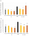
Percentage variation vs. 0 h of skin redness in the placebo (green line), 100 mg (orange line), and 250 mg (blue line) Nutroxsun® (Nx) treatment groups. Results are shown for the global panel (a) and the subgroup of subjects with light phototypes (b). formula image product intake. Statistically significant vs. placebo is reported as * p < 0.05 and ** p < 0.01.
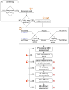
Figure 7
Figure 7
Study flow and design. Subjects who met the inclusion criteria were then screened by a dermatologist. During the screening visit, a physical examination was carried out to assess the uniformity of the test area (back). Subjects meeting the inclusion criteria were then enrolled and randomized to participate in the study. Legends: formula image information, formula image physical examination, formula image informed consent signature, formula image eligibility check, formula image randomization, R skin redness, formula image product intake.

References

    1. Holick M.F. Sunlight and Vitamin D for Bone Health and Prevention of Autoimmune Diseases, Cancers, and Cardiovascular Disease. Am. J. Clin. Nutr. 2004;80:1678S–1688S. doi: 10.1093/ajcn/80.6.1678S. - DOI - PubMed
    1. Weller R.B. Sunlight Has Cardiovascular Benefits Independently of Vitamin D. Blood Purif. 2016;41:130–134. doi: 10.1159/000441266. - DOI - PubMed
    1. Passeron T., Krutmann J., Andersen M., Katta R., Zouboulis C. Clinical and Biological Impact of the Exposome on the Skin. J. Eur. Acad. Dermatol. Venereol. 2020;34:4–25. doi: 10.1111/jdv.16614. - DOI - PubMed
    1. Greaves M.W., Sondergaard J. Pharmacologic Agents Released in Ultraviolet Inflammation Studied by Continuous Skin Pefusion. J. Investig. Dermatol. 1970;54:365–367. doi: 10.1111/1523-1747.ep12259058. - DOI - PubMed
    1. Benrath J., Eschenfelder C., Zimmerman M., Gillardon F. Calcitonin Gene-Related Peptide, Substance P and Nitric Oxide Are Involved in Cutaneous Inflammation Following Ultraviolet Irradiation. Eur. J. Pharmacol. 1995;293:87–96. doi: 10.1016/0926-6917(95)90022-5. - DOI - PubMed

Publication types

MeSH terms