. 2025 Apr 25;26(9):4055.
doi: 10.3390/ijms26094055.

Identification of Potential Prophylactic Medical Countermeasures Against Acute Radiation Syndrome (ARS)

Affiliations

Identification of Potential Prophylactic Medical Countermeasures Against Acute Radiation Syndrome (ARS)

Kia T Liermann-Wooldrik et al. Int J Mol Sci. .

Abstract

Acute radiation syndrome (ARS) occurs when hematopoietic or gastrointestinal cells are damaged by radiation exposure causing DNA damage to the bone marrow and gastrointestinal epithelial stem cell populations. In these highly proliferative cell types, DNA damage inhibits stem cell repopulation. In humans and animals, this inability to regenerate stem cells is lethal. Within this manuscript, several compounds, Amifostine, Captopril, Ciprofloxacin, PrC-210, 5-AED (5-androstene-3β,17β-diol), and 5-AET (5-androstene-3β,7β,17B-triol), are assessed for their ability to protect against ARS in an in vitro and/or in vivo setting. ARS was accomplished by irradiating mouse bone marrow cells or rat intestinal epithelial (IEC-6) cells in vitro with 4-8 Gy and in vivo by exposing Mus musculus to 7.3 Gy of whole-body irradiation. The primary endpoints of this study include cellular viability, DNA damage via γ-H2AX, colony formation, and overall survival at 30-days post-irradiation. In addition to evaluating the radioprotective performance of each compound, this study establishes a distinct set of in vitro assays to predict the overall efficacy of potential radioprotectors in an in vivo model of ARS. Furthermore, these results highlight the need for FDA-approved medical intervention to protect against ARS.

Keywords: DNA damage; acute radiation syndrome; radioprotection.


Conflict of interest statement

The authors declare no conflicts of interest.

Figures

Figure 1
Figure 1
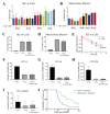
Amifostine protects rat intestinal cells and mouse bone marrow cells from acute radiation damage. (A) Amifostine (Amif) was administered to IEC-6 at doses of 100 and 1000 µM 30 min before exposure to 6 Gy of radiation, and cell death enumerated. (B) Cell death was quantified by viability in mouse bone marrow cells treated with various doses of Amif and 6 Gy of radiation. (C) IEC-6 cells and (D) mouse bone marrow cells were treated with 100 µM of Amif and 5 Gy of radiation to determine the accumulation of DNA damage via γ-H2AX staining. (E) A clonogenic assay of IEC-6 cells pre-treated with 1 mM of Amif 30 min before radiation was used to determine the ability of these cells to form colonies. (F) Colony formation units of granulocytes (CFU-G); (G) macrophages (CFU-M); (H) granulocyte, macrophage (CFU-GM); and (I) granulocyte, erythrocyte, macrophage, and megakaryocyte (CFU-GEMM) progenitors were assayed in mouse bone marrow cells treated with 100 µM of Amif 30 min before receiving 4 Gy of radiation. All experiments were executed three times. * Denotes a significant difference (p ≤ 0.05) as compared to the untreated, irradiated control.
Figure 2
Figure 2
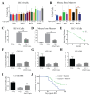
Ciprofloxacin does not protect rat intestinal cells and mouse bone marrow cells from acute radiation damage. (A) Quantification of cell death was observed in IEC-6 cells treated with 5, 10, or 25 µM of Ciprofloxacin (Cipro) one hour before exposure to 4, 6, or 8 Gy. (B) Cell death was quantified in mouse bone marrow cells treated with various concentrations of Cipro. (C) DNA damage was assessed by enumerating γ-H2AX+ IEC-6 cells or (D) mouse bone marrow cells treated with 25 µM of Cipro. (E) Clonogenic capacity was measured after administering 25 µM of Cipro to IEC-6 cells one hour before radiation. (F) Colony formation units of granulocytes (CFU-G); (G) macrophages (CFU-M); (H) granulocyte, macrophage (CFU-GM); and (I) granulocyte, erythrocyte, macrophage, and megakaryocyte (CFU-GEMM) progenitors were quantified in mouse bone marrow cells treated with Cipro (25 µM) and 4 Gy. Three replicates were completed from each assay. (J) Mice were administered 0.67 mg/mL of Cipro 1 h before whole-body irradiation (7.3 Gy) over 14 days after radiation. The overall 30-day survival was not significantly enhanced by Cipro treatment. Twenty mice per group were used to evaluate survival.
Figure 3
Figure 3
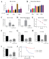
Captopril protects against ARS. (A). Toxicity and protection from radiation-induced cell death was evaluated in IEC-6 cells treated with 50, 100, or 400 µM one hour before exposure to 4, 6, or 8 Gy. (B) The percentage of dead cells was quantified in mouse bone marrow cells treated with Captopril (Capto, 400 µM). (C) DNA damage was quantified by enumerating γ-H2AX+ IEC-6 cells and (D) mouse bone marrow cells pre-treated with 400 µM of Capto and exposed to 5 Gy. (E) A clonogenic survival assay was completed on IEC-6 cells dosed with Capto (400 µM), or the control (0 µM), and radiation. To assay the colony formation of bone marrow cells, 400 µM of Capto was administered to the cells 1 h before radiation (4 Gy). (F) Colony formation units of granulocytes (CFU-G); (G) macrophages (CFU-M); (H) granulocyte, macrophage (CFU-GM); and (I) granulocyte, erythrocyte, macrophage, and megakaryocyte (CFU-GEMM) progenitors were quantified 10 days after drug and radiation treatment. Three replicates were completed from each assay. * Indicates a significant difference (p ≤ 0.05) as compared to the untreated, irradiated control. (J) Mice were administered 110 mg/mL of Capto 48, 24, and 1 h before whole-body irradiation (7.3 Gy). The overall 30-day survival was significantly enhanced by Capto treatment. Forty mice per group were used to evaluate survival.
Figure 4
Figure 4
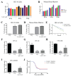
PrC-210 protects against ARS both in vitro and in vivo. (A) The viability of IEC-6 cells and (B) mouse bone marrow cells treated with 0.5, 1, or 2 mg/mL thirty minutes before exposure to 6 or 8 Gy was assessed as the percentage of dead cells. (C) DNA damage, indicated by γ-H2AX staining, in IEC-6 cells and (D) mouse bone marrow cells, pre-treated with 1 mg/mL of PrC-210 before exposure to 5 Gy. (E) A total of 1 mg/mL of PrC-210, or control, was administered to IEC-6 cells prior to 4, 6, and 8 Gy of radiation exposure when compared to assessments of the control and colony formation. To assay the colony formation of bone marrow cells, 1 mg/mL of PrC-210 was administered to the cells 30 min before radiation (4 Gy). (F) Colony formation units of granulocytes (CFU-G); (G) macrophages (CFU-M); (H) granulocyte, macrophage (CFU-GM); and (I) granulocyte, erythrocyte, macrophage, and megakaryocyte (CFU-GEMM) progenitors were quantified in mouse bone marrow cells treated with PrC-210 (1 mg/mL) prior to radiation exposure (4 Gy). Three biological replicates were completed from each assay. (J) Mice were administered 900 mg/kg of PrC-210 30 min before whole-body irradiation (7.3 Gy). The overall 30-day survival was significantly enhanced by PrC-210 treatment. Thirty-five mice per group were used to evaluate survival. * Indicates a significant difference (p ≤ 0.05) compared to the untreated, irradiated control.
Figure 5
Figure 5
5-AED is a radioprotector in vitro and in vivo. (A) Radiation-induced cell death in IEC-6 cells treated with 0, 0.5, or 1 µM of 5-AED one hour before exposure to 6 or 8 Gy. (B) Quantification of cell death in mouse bone marrow cells treated with 5-AED. (C) DNA damage in IEC-6 cells treated with or without 5-AED (0.5 µM) and 5 Gy of radiation was assessed by enumerating the percentage of cells positive for γ-H2AX staining. (D) Percentage of γ-H2AX+ mouse bone marrow cells treated with or without 0.5 µM of 5-AED and 5 Gy. (E) Clonogenic capacity of IEC-6 cells was evaluated at 4, 6, and 8 Gy when treated with control (PEG-Methanol) or 0.5 µM of 5-AED. (F) Colony formation units of granulocytes (CFU-G); (G) macrophages (CFU-M); (H) granulocyte, macrophage (CFU-GM); and (I) granulocyte, erythrocyte, macrophage, and megakaryocyte (CFU-GEMM) progenitors were quantified 10 days after bone marrow cells were treated with 5-AED (0.5 µM) one hour prior to radiation exposure (4 Gy). Three biological replicates were completed from each assay. * Indicates a significant difference (p ≤ 0.05) from the untreated, irradiated control. (J) Mice were administered 30 mg/kg of 5-AED 24 h before whole-body irradiation (7.3 Gy). 5-AED treatment significantly protected the overall 30-day survival. Twenty mice per group were used to evaluate survival.
Figure 6
Figure 6
5-AET does not protect against radiation damage. (A) Radiation-induced cell death in IEC-6 cells treated with 0, 0.5, 1, or 2 µM of 5-AET one hour before exposure to 6 or 8 Gy. (B) Quantification of cell death in mouse bone marrow cells treated with 5-AET. (C) DNA damage in IEC-6 cells treated with or without 5-AET (0.5 µM) and 5 Gy of radiation was assessed by enumerating the percentage of cells positive for γ-H2AX staining. (D) Percentage of γ-H2AX+ mouse bone marrow cells treated with or without 0.5 µM of 5-AET and 5 Gy. (E) Clonogenic capacity of IEC-6 cells was evaluated at 4, 6, and 8 Gy when treated with control (PEG-Methanol) or 0.5 µM of 5-AET. (F) Colony formation units of granulocytes (CFU-G); (G) macrophages (CFU-M); (H) granulocyte, macrophage (CFU-GM); and (I) granulocyte, erythrocyte, macrophage, and megakaryocyte (CFU-GEMM) progenitors were quantified 10 days after bone marrow cells were treated with 5-AET (1 µM) one hour prior to radiation exposure (4 Gy). Three biological replicates were completed from each assay. * Indicates a significant difference (p ≤ 0.05) from the untreated, irradiated control. (J) Mice were administered 30 mg/kg of 5-AET 24 h before whole-body irradiation (7.3 Gy). 5-AET treatment did not significantly alter the overall 30-day survival. Twenty mice per group were used to evaluate survival.
Figure 7
Figure 7
Predictive capacity of in vitro experiments. Comparison of the number of in vitro assays showing radioprotection versus the in vivo 30-day percent survival. Captopril and 5-AED are the two outlier compounds. R2 = 0.7115.

References

    1. Hall E.J., Giaccia A.J. Radiobiology for the Radiologist. 6th ed. Lippincott Williams and Wilkins; Philadellphia, PA, USA: 2006.
    1. Du C., Gao Z., Venkatesha V.A., Kalen A.L., Chaudhuri L., Spitz D.R., Cullen J.J., Oberley L.W., Goswami P.C. Mitochondrial ROS and radiation induced transformation in mouse embryonic fibroblasts. Cancer Biol. Ther. 2009;8:1962–1971. doi: 10.4161/cbt.8.20.9648. - DOI - PMC - PubMed
    1. Indo H.P., Inanami O., Koumura T., Suenaga S., Yen H.-C., Kakinuma S., Matsumoto K.-I., Nakanishi I., Clair W.S., Clair D.K.S., et al. Roles of mitochondria-generated reactive oxygen species on X-ray-induced apoptosis in a human hepatocellular carcinoma cell line, HLE. Free Radic. Res. 2012;46:1029–1043. doi: 10.3109/10715762.2012.698012. - DOI - PubMed
    1. Riley P.A. Free radicals in biology: Oxidative stress and the effects of ionizing radiation. Int. J. Radiat. Biol. 1994;65:27–33. doi: 10.1080/09553009414550041. - DOI - PubMed
    1. Yamaguchi M., Kashiwakura I. Role of reactive oxygen species in the radiation response of human hematopoietic stem/progenitor cells. PLoS ONE. 2013;8:e70503. doi: 10.1371/journal.pone.0070503. - DOI - PMC - PubMed

MeSH terms

LinkOut - more resources