. 2025 Apr 29;26(9):4208.
doi: 10.3390/ijms26094208.

In Vitro Immunomodulatory Effects of Equine Adipose Tissue-Derived Mesenchymal Stem Cells Primed with a Cannabidiol-Rich Extract

Affiliations

In Vitro Immunomodulatory Effects of Equine Adipose Tissue-Derived Mesenchymal Stem Cells Primed with a Cannabidiol-Rich Extract

Lorena Battistin et al. Int J Mol Sci. .

Abstract

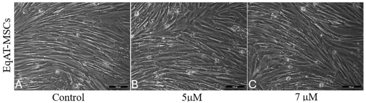
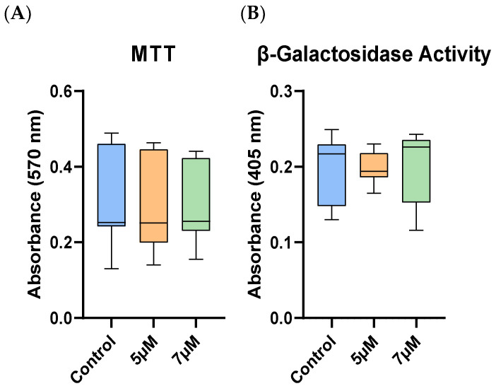
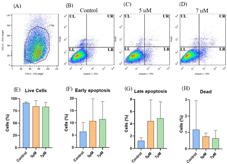
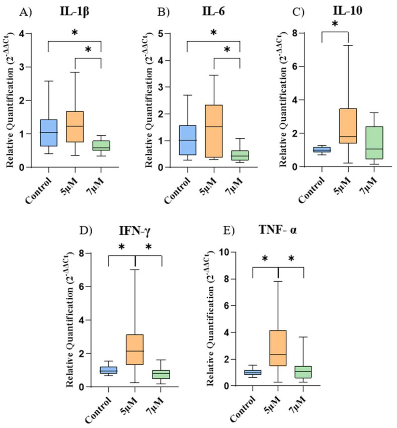
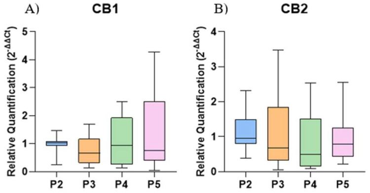
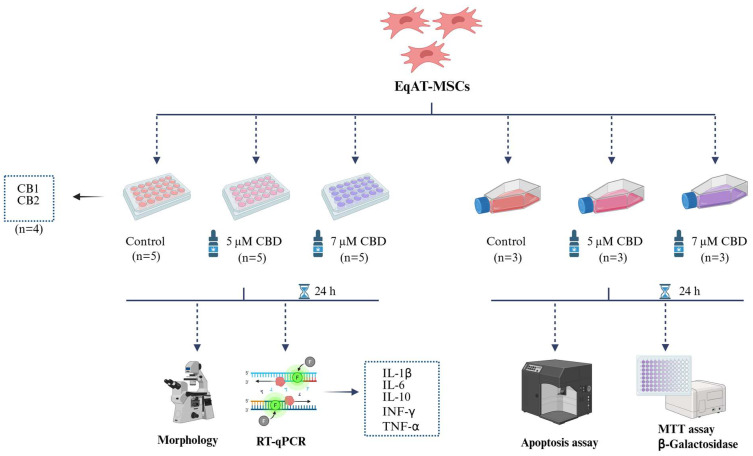
Cell-based therapy using mesenchymal stem cells (MSCs) shows promise for treating several diseases due to their anti-inflammatory and immunomodulatory properties. To enhance the therapeutic potential of MSCs, in vitro priming strategies have been explored. Cannabidiol (CBD), a non-psychoactive compound derived from cannabis, may influence MSC proliferation, differentiation, and immunomodulatory properties. This study evaluates the immunomodulatory potential of equine adipose tissue-derived MSCs (EqAT-MSCs) primed with a CBD-rich cannabis extract. EqAT-MSCs (P3) were primed with CBD concentrations of 5 µM and 7 µM for 24 h. Morphological analysis, MTT assay, β-galactosidase activity, apoptosis assays, and gene expression of interleukins IL-1β, IL-6, IL-10, interferon-gamma (IFN-γ), and tumor necrosis factor-alpha (TNF-α) were conducted. Additionally, cannabinoid receptor 1 (CB1) and 2 (CB2) expression were evaluated in naïve EqAT-MSCs (P2-P5). The naïve EqAT-MSCs expressed CB1 and CB2 receptors. Priming with 5 µM significantly increased the expression of IL-10, TNF-α, and IFN-γ, while 7 µM decreased IL-1β and IL-6 expression. No significant changes were observed in other cytokines, MTT, β-galactosidase activity, or apoptosis. These findings demonstrate that naïve EqAT-MSCs express CB1 and CB2 receptors and priming with the extract modulates the expression of pro- and anti-inflammatory cytokines, highlighting its potential immunomodulatory role in EqAT-MSC-based therapies.

Keywords: cannabinoid receptors; cell therapy; cytokines; horse; phytocannabinoid.


Conflict of interest statement

The authors declare no conflicts of interest.

Figures

Figure 1
Figure 1
Morphological characterization of equine adipose tissue-derived mesenchymal stem cells (EqAT-MSCs). (A) Control EqAT-MSCs cultured in complete culture medium; (B) EqAT-MSCs primed with Cannabidiol (CBD)-rich cannabis extract at concentrations of 5 μM and (C) 7 μM. No morphological changes were observed compared to the control group. Magnification, ×200. Scale bar = 100 μM.
Figure 2
Figure 2
Cellular metabolic and β-galactosidase activities in equine adipose tissue-derived mesenchymal stem cells (EqAT-MSCs). No significant differences were observed after priming with cannabidiol (CBD)-rich cannabis extract in (A) cellular metabolic activity (MTT assay) and (B) senescence-associated β-galactosidase activity. Data are shown as medians, interquartile ranges, minimum, and maximum values. Kruskal–Wallis test.
Figure 3
Figure 3
Apoptosis assay (Annexin V × 7-AAD): (AD) Representative gating strategy for the analysis of cell viability using Annexin-V FITC and 7-AAD. (A) Selection of the cellular population. (B) Control group. (C) Equine adipose tissue-derived mesenchymal stem cells (EqAT-MSCs) primed with cannabidiol (CBD)-rich cannabis extract at concentrations of 5 μM (D) and 7 μM. Quadrants are labeled as follows: UL (Upper Left) represents dead cells, LL (Lower Left) represents live cells, UR (Upper Right) represents late apoptotic cells, and LR (Lower Right) represents early apoptotic cells. No significant changes were observed when comparing (E) the live cells, (F) early apoptotic cells, (G) late apoptotic cells, and (H) dead cells of the control group with EqAT-MSCs primed with CBD-rich cannabis extract at concentrations of 5 μM and 7 μM. Data are represented as the mean ± standard deviation, one-way analysis of variance (ANOVA).
Figure 4
Figure 4
Relative expression of the cytokines among the experimental groups. (A) Interleukin 1 beta (IL-1β), (B) interleukin-6 (IL-6), (C) interleukin-10 (IL-10), (D) interferon gamma (IFN-γ), and (E) tumor necrosis factor-alpha (TNF-α). Data are represented as medians, interquartile ranges, and minimum and maximum values (* p < 0.05). Kruskal–Wallis and Dunn’s tests.
Figure 5
Figure 5
Relative expression of cannabinoid receptors 1 (CB1) and 2 (CB2) in the control group of naïve equine adipose tissue-derived mesenchymal stem cells (EqAT-MSCs) across different passages (P2–P5): (A) CB1. (B) CB2. No significant difference was observed between the passages. Data are represented as medians, interquartile ranges, minimum, and maximum values. Kruskal–Wallis test.
Figure 6
Figure 6
Experimental design. Equine adipose tissue-derived mesenchymal stem cells (EqAT-MSCs) (P3) were primed with 5 µM of cannabidiol (CBD)-rich cannabis extract + complete culture medium and the other group was primed with 7 µM of CBD-rich cannabis extract + complete culture medium. EqAT-MSCs cultured in a complete culture medium were used as the control. After 24 h of priming, the morphological evaluation and gene expression analysis of cytokines such as interleukin 1 beta (IL-1β), interleukin 6 (IL-6), interleukin 10 (IL-10), interferon-gamma (IFN-γ), and tumor necrosis factor-alpha (TNF-α) were performed. Cell metabolic activity was assessed using the MTT assay, and cellular senescence was evaluated through β-galactosidase activity. The apoptosis assay was carried out using annexin and 7-AAD. In addition to the evaluation of cannabinoid receptors 1 (CB1) and 2 (CB2) gene expression in naïve EqAT-MSCs (P2–P5). Created in BioRender. (https://BioRender.com/q24g045 (accessed on 23 April 2025)).

Similar articles

References

    1. Andrzejewska A., Dabrowska S., Lukomska B., Janowski M. Mesenchymal stem cells for neurological disorders. Adv. Sci. 2021;8:2002944. doi: 10.1002/advs.202002944. - DOI - PMC - PubMed
    1. Fan X.L., Zhang Y., Li X., Fu Q.L. Mechanisms underlying the protective effects of mesenchymal stem cell-based therapy. Cell. Mol. Life Sci. 2020;77:2771–2794. doi: 10.1007/s00018-020-03454-6. - DOI - PMC - PubMed
    1. Alvites R., Branquinho M., Sousa A.C., Lopes B., Sousa P., Maurício A.C. Mesenchymal stem/stromal cells and their paracrine activity—Immunomodulation mechanisms and how to influence the therapeutic potential. Pharmaceutics. 2022;14:381. doi: 10.3390/pharmaceutics14020381. - DOI - PMC - PubMed
    1. Chen P.M., Liu K.J., Hsu P.J., Wei C.F., Bai C.H., Ho L.J., Sytwu H.K., Yen B.L. Induction of immunomodulatory monocytes by human mesenchymal stem cell-derived hepatocyte growth factor through ERK1/2. J. Leukoc. Biol. 2014;96:295–303. doi: 10.1189/jlb.3A0513-242R. - DOI - PubMed
    1. Maličev E., Jazbec K. An overview of mesenchymal stem cell heterogeneity and concentration. Pharmaceuticals. 2024;17:350. doi: 10.3390/ph17030350. - DOI - PMC - PubMed

MeSH terms

LinkOut - more resources