Review
. 2025 May 6;26(9):4416.
doi: 10.3390/ijms26094416.

Antibacterial Activity of the p53 Tumor Suppressor Protein-How Strong Is the Evidence?

Affiliations
Review

Antibacterial Activity of the p53 Tumor Suppressor Protein-How Strong Is the Evidence?

Agnieszka Gdowicz-Kłosok et al. Int J Mol Sci. .

Abstract

The p53 tumor suppressor is best known for controlling the cell cycle, apoptosis, DNA repair, and metabolism, but it also regulates immunity and is able to impede the live cycle of viruses. For this reason, these infectious agents encode proteins which inactivate p53. However, what is less known is that p53 can also be inactivated by human pathogenic bacteria. It is probably not due to collateral damage, but specific targeting, because p53 could interfere with their multiplication. The mechanisms of the antibacterial activity of p53 are poorly known. However, they can be inferred from the results of high-throughput studies, which have identified more than a thousand p53-activated genes. As it turns out, many of these genes code proteins which have proven or plausible antibacterial functions like the efficient detection of bacteria by pattern recognition receptors, the induction of pro-inflammatory pyroptosis, the recruitment of immune cells, direct bactericidal activity, and the presentation of bacterial metabolites to lymphocytes. Probably there are more antibacterial, p53-regulated functions which were overlooked because laboratory animals are kept in sterile conditions. In this review, we present the outlines of some intriguing antibacterial mechanisms of p53 which await further exploration. Definitely, this area of research deserves more attention, especially in light of the appearance of antibiotic-resistant bacterial strains.

Keywords: Helicobacter pylori; MDM2; actinomycin D; defensin; innate immunity; nutlin-3a; tuberculosis.


Conflict of interest statement

The authors declare no conflicts of interest.

Figures

Figure 1
Figure 1
Bacteria antagonize p53 protein by mechanisms of its destabilization or by reducing expression of its mRNA. Bacteria produce proteins (bps), which interact with cellular molecules interfering with the p53 pathway. For instance, one of the proteins (IpgD) produced by S. flexneri promotes activation of AKT kinase, which phosphorylates and activates MDM2, which is a major negative regulator of p53 inducing its degradation. Another S. flexneri protein (VirA) induces proteolysis of calpastatin, which is a negative regulator of calpain. Liberated calpain snips off the N-terminus of p53. Other bacteria also act through the activation of AKT. For instance, in H. pylori, this function is played by the CagA protein. On the other hand, C. trachomatis activates EGFR (epidermal growth factor receptor), which by its natural signaling pathway stimulates AKT. N. gonorrhoeae arrests the cell cycle and induces DNA breaks in host cells, which in normal conditions would activate p53, but infection with this bacterium, by an unknown mechanism, reduces expression of p53 mRNA. K. pneumoniae activates Toll-like receptor 4 (TLR4), which indirectly activates the NF-κB pathway, which through mediators causes the destabilization of p53 mRNA. The only known bacterium that apparently activates p53 is Salmonella Typhimurium, which through its AvrA protein promotes p53 acetylation, which is associated with its stabilization and activation.
Figure 2
Figure 2
The p53 activates at least three genes, which encode the components of inflammasomes. A simplified model of pro-inflammatory caspase-1 activation. Inflammasomes are multiprotein complexes promoting inflammation. They consist of pro-caspase-1 (localized centrally) linked to various pattern recognition receptors (PRRs, localized peripherally) by the ASC protein. Pro-caspase-1 and ASC are linked through CARD domains, whereas ASC and PRRs are linked through PYRIN domains. Binding of a ligand (e.g., a bacterial molecule) to a PRR induces a series of molecular alterations, which lead to the cleavage of pro-caspase-1 into p10 and p20 domains, which as a dimer of dimers form active-caspase-1. This enzyme cleaves inactive pro-inflammatory IL-1β and IL-18 into active molecules. Moreover, caspase-1 cuts gasdermin proteins, whose N-terminal fragments form pores within the plasma membrane. These pores are used by active cytokines to escape into extracellular space. Moreover, the pores contribute to cell swelling and death, which is called pyroptosis. Interestingly, NLRP1 can directly interact with pro-caspase-1 through the CARD domains of both proteins. The NLRP1 inflammasome is poorly studied, but it can be the predominant inflammasome in human barrier cells. The genes coding these components of the inflammasomes (pro-caspase-1, NLRP1, and NLRP3) are directly activated by p53. Moreover, p53 activates a gene for one of the gasdermins—GSDME.
Figure 3
Figure 3
Multiple roles of p53 in antibacterial defense. The genes regulated by p53 involved in the indicated functions and described in this review are shown in circles. The “+” sign means that these genes are up-regulated in a p53-dependent fashion. There are probably more antibacterial genes regulated by p53 that have not been identified yet, hence the question mark.
Figure 4
Figure 4
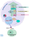
The antibacterial role of p53, which is mediated by up-regulation of the TNFRSF14 gene. This gene codes the cell surface protein, which can either act as a ligand or as a receptor depending on the cellular context. In this example, TNFRSF14 protein acts as a receptor for CD160 protein expressed on innate-like intraepithelial lymphocytes. Stimulation of the receptor by CD160 protein triggers a cascade of events involving the activation of NIK kinase (NF-κB-inducing kinase), which phosphorylates and activates the STAT3 protein. This transcription factor induces a plethora of host defense genes, e.g., the gene for β-defensin 3, the genes for pro-inflammatory cytokines and chemokines.

References

    1. Levine A.J. The many faces of p53: Something for everyone. J. Mol. Cell. Biol. 2019;11:524–530. doi: 10.1093/jmcb/mjz026. - DOI - PMC - PubMed
    1. Nagata J., Kijima H., Takagi A., Ito M., Goto K., Yamazaki H., Nakamura M., Mine T., Ueyama Y. Helicobacter pylori induces chronic active gastritis in p53-knockout mice. Int. J. Mol. Med. 2004;13:773–777. doi: 10.3892/ijmm.13.6.773. - DOI - PubMed
    1. Carlsen L., Zhang S., Tian X., De La Cruz A., George A., Arnoff T.E., El-Deiry W.S. The role of p53 in anti-tumor immunity and response to immunotherapy. Front. Mol. Biosci. 2023;10:1148389. doi: 10.3389/fmolb.2023.1148389. - DOI - PMC - PubMed
    1. Łasut-Szyszka B., Rusin M. The Wheel of p53 Helps to Drive the Immune System. Int. J. Mol. Sci. 2023;24:7645. doi: 10.3390/ijms24087645. - DOI - PMC - PubMed
    1. Łasut-Szyszka B., Gdowicz-Kłosok A., Małachowska B., Krześniak M., Będzińska A., Gawin M., Pietrowska M., Rusin M. Transcriptomic and proteomic study of cancer cell lines exposed to actinomycin D and nutlin-3a reveals numerous, novel candidates for p53-regulated genes. Chem. Biol. Interact. 2024;392:110946. doi: 10.1016/j.cbi.2024.110946. - DOI - PubMed

MeSH terms

Substances