. 2025 Apr 25;17(9):1445.
doi: 10.3390/nu17091445.

(-)-Oleuropein as a Novel Metastatic Castration-Resistant Prostate Cancer Progression and Recurrence Suppressor via Targeting PCSK9-LDLR Axis

Affiliations

(-)-Oleuropein as a Novel Metastatic Castration-Resistant Prostate Cancer Progression and Recurrence Suppressor via Targeting PCSK9-LDLR Axis

Nehal A Ahmed et al. Nutrients. .

Abstract

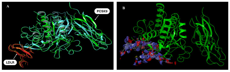
Background/Objectives: Prostate cancer (PC) is among the most common malignancy in men. Several newly diagnosed patients have a locally advanced disease and distant metastasis at the initial diagnosis time. Castration-resistant PC (CRPC) patients have 100% recurrence incidence despite completing a therapeutic regimen, leading to high mortality. Androgen deprivation therapy and androgen inhibitors are initially effective, but resistance is inevitably developed. Epidemiological studies indicated that the Mediterranean diet, with high olive phenolic contents, is associated with a lower incidence of certain malignancies. This study aims at exploring the mCRPC progression and recurrence-suppressive and molecular effects of the major olive leaf phenolic glucoside (-)-oleuropein (OLE). Results: OLE downregulated the levels of proprotein convertase subtlisin/klexin type 9 (PCSK9) and normalized the low-density lipoprotein receptor (LDLR) in PC cells in vitro. Thus, a PCSK9-LDLR protein-protein interaction (PPI) in silico model was generated and used to assess OLE and its aglycone (OA) ability to bind at PCSK9 and thereby interfere with PCSK9-LDLR PPI. OLE perfectly filled the PCSK9 interface versus OA. Both OLE and OA showed virtual potential to interfere with PCSK9-LDLR PPI. OLE showed modest in vitro viability, migration, and clonogenicity suppressive effects on diverse human PC cell lines. OLE effectively suppressed mCRPC progression and recurrence in a nude mouse xenograft model. RNA-sequencing results proved the PCSK1, PCSK2, and PCSK9 downregulation in OLE-treated recurrent tumors versus vehicle control. Conclusions: Oleuropein is a novel lead useful for the control of mCRPC progression and the prevention of its recurrence via targeting PCSK9 expression and PPI with LDLR.

Keywords: PCSK9-LDLR; metastatic castration-resistant prostate cancer; oleuropein; olive phenolics; protein–protein interaction; recurrence.


Conflict of interest statement

The authors declare no conflicts of interest.

Figures

Figure 1
Figure 1
Favorable predicted molecular modeling PCSK9-LDLR interactions at various sites (AG). Hydrogen bonds are shown as yellow dotted lines, amino acid residues interacting on the PCSK9 interface and LDLR are shown in green.
Figure 2
Figure 2
PCSK9-LDLR generated model protein alignment. (A) Overlay of docked pose of PCSK9 (green)-LDLR (orange) using the PDB available crystal structure 3BPS PCSK9 (blue)-LDLR (red). (B) Possible binding site detection at PCSK9 using SiteMap.
Figure 3
Figure 3
Molecular docking of OLE at the PCSK9-LDLR interface. (A) OLE showed favorable hydrogen bonding with the PCSK9 at the amino acid residues Asp374, Arg194, Phe379, and Ser153. Red arrows indicate critical amino acids engaged in binding interactions. (B) Redocking of PCSK9-OLE complex with LDLR, the presence OLE at the PCSK9 hindered the specific docking of EGF-A of LDLR at this site and resulted in an improper docking pose.
Figure 3
Figure 3
Molecular docking of OLE at the PCSK9-LDLR interface. (A) OLE showed favorable hydrogen bonding with the PCSK9 at the amino acid residues Asp374, Arg194, Phe379, and Ser153. Red arrows indicate critical amino acids engaged in binding interactions. (B) Redocking of PCSK9-OLE complex with LDLR, the presence OLE at the PCSK9 hindered the specific docking of EGF-A of LDLR at this site and resulted in an improper docking pose.
Figure 4
Figure 4
Molecular docking of OA at the PCSK9 interface. (A) OA showed reduced possibility to fill in the shallow interface at the PCSK9, only showing hydrogen bonding with the amino acid residues Asp374, Arg194, and Phe379. Red arrows indicate critical amino acids engaged in binding interactions. (B) Redocking PCSK9-OA complex with LDLR suggests that the binding of OA at PCSK9 hindered its docking ability with LDLR EGF-A.
Figure 5
Figure 5
In vitro effects of OLE on the viability of the human non-tumorigenic prostate cells and diverse PC cell lines determined by MTT assay. (A) Effects of OLE on the viability of the human non-tumorigenic RWPE-1 cells. (BE) Effects of OLE on the viability of the human PC cell lines CWR-R1ca, DU-145, PC-3, and LNCaP, respectively. ns: Non-statistical significance at p > 0.05. * Statistical significance at p < 0.05, ** statistical significance at p < 0.01, and *** statistical significance at p < 0.001.
Figure 6
Figure 6
Anti-migratory and colony formation suppressive effects of OLE treatments against the migration and colonization of CWR-R1ca, DU-145, PC-3, and LNCaP PC cells. (A) Wound-healing assay images captured after 24 h incubation of CWR-R1ca, DU-145, PC-3, and LNCaP cells with OLE or VC. (B) Quantitative analysis of the percentage of migration (wound closure) with various OLE treatment doses. Vertical bars indicate the percentage of wound closure of cells after 24 h of wound scratching, calculated relative to the wound distance at 0 h in each treatment group. (C) Representative images of colony formation of CWR-R1ca DU-145, PC-3, and LNCaP PC cells treated with OLE treatments over 12 days and finally stained with Giemsa stain at the end of the experiment. (D) Quantification of OLE colony formation suppressive effects. Vertical bars indicate the percentage of colony formation relative to vehicle control. * p < 0.05, ** p < 0.01, and *** p < 0.001, indicate statistical significance compared to their respective vehicle-treated controls.
Figure 7
Figure 7
In vitro effects of OLE treatments on the expression levels of PCSK9 and LDLR in CWR-R1ca mCRPC cell line. (A) Western blots of PCSK9 and LDLR expression levels in cultured CWR-R1ca cells subjected to 25, 50, and 100 µM of OLE treatments in comparison to VC-treated cells. (B) Densitometric analysis of PCSK9 and LDLR expression and the integrated optical density of each band normalized with the corresponding density found for β-tubulin in the same blot. Vertical bars in the graph indicate the normalized integrated optical density of bands visualized in each lane. * p < 0.05 and ** p < 0.01, and *** p < 0.001 indicate statistical significance compared to their respective vehicle-treated controls.
Figure 8
Figure 8
In vivo mCRPC CWR-R1ca-Luc progression suppressive effect of OLE 75 mg/kg, ip 3×/week, versus VC in a nude mouse xenograft model. (A) Photographic comparison of surgically excised primary tumors. Top row shows VC-treated primary tumors. Bottom row OLE-treated primary tumors. (B) Comparative monitoring of tumors volume in OLE and VC-treated groups over the dosing period. (C) Comparison of OLE and VC-treated tumor weights. (D) Western blotting image comparison of the effects of OLE versus VC treatments on the expression of PCSK9 and LDLR in collected CWR-R1ca-Luc primary tumors. (E) Densitometric analysis of PCSK9 and LDLR expression levels in primary tumors treated with OLE versus vehicle controls. Vertical bars in the graph indicate the normalized integrated optical density of bands visualized in each lane. * p < 0.05 and ** p < 0.01 indicates statistical significance compared to their respective vehicle-treated controls.
Figure 9
Figure 9
In vivo mCRPC recurrence suppressive effect of OLE 75 mg/kg, ip, 3×/week versus VC in a nude mouse xenograft model after surgical excision of primary tumors. (A) Whole animal bioluminescence imaging at the end of the study comparing OLE locoregional recurrence suppressive effects versus VC treatment effects. (B) Bioluminescence images of the collected organs (liver, lung, brain bone, kidney) on the last day of the study. (C) Mice body weight monitoring throughout the study period. (D,E) Comparison of OLE versus VC-treated mean tumor volume and weight collected after animals were sacrificed. (F) Western blots and densitometric analysis of PCSK9 and LDLR expression levels in OLE versus VC-treated nude mouse livers at the end of the study. Vertical bars in the graph indicate the normalized integrated optical density of bands visualized in each lane. ns indicates statistical non-significance, * p < 0.05, ** p < 0.01, and *** p < 0.001 indicate statistical significance compared to their respective vehicle-treated controls.

References

    1. Siegel R.L., Kratzer T.B., Giaquinto A.N., Sung H., Jemal A. Cancer statistics, 2025. CA Cancer J. Clin. 2025;75:10–45. doi: 10.3322/caac.21871. - DOI - PMC - PubMed
    1. Wang G., Zhao D., Spring D.J., DePinho R.A. Genetics and biology of prostate cancer. Genes Dev. 2018;32:1105–1140. doi: 10.1101/gad.315739.118. - DOI - PMC - PubMed
    1. Henríquez I., Roach M., 3rd, Morgan T.M., Bossi A., Gómez J.A., Abuchaibe O., Couñago F. Current and emerging therapies for metastatic castration-resistant prostate cancer (mCRPC) Biomedicines. 2021;9:1247. doi: 10.3390/biomedicines9091247. - DOI - PMC - PubMed
    1. Giacinti S., Poti G., Roberto M., Macrini S., Bassanelli M., DI Pietro F., Aschelter A.M., Ceribelli A., Ruggeri E.M., Marchetti P. Molecular basis of drug resistance and insights for new treatment approaches in mCRPC. Anticancer. Res. 2018;38:6029–6039. doi: 10.21873/anticanres.12953. - DOI - PubMed
    1. Go G.W., Mani A. Low-density lipoprotein receptor (LDLR) family orchestrates cholesterol homeostasis. Yale J. Biol. Med. 2012;85:19–28. - PMC - PubMed

MeSH terms

LinkOut - more resources