. 2025 May 3;18(9):2099.
doi: 10.3390/ma18092099.

Critical Design Parameters of Tantalum-Based Comb Structures to Manipulate Mammalian Cell Morphology

Affiliations

Critical Design Parameters of Tantalum-Based Comb Structures to Manipulate Mammalian Cell Morphology

Hassan I Moussa et al. Materials (Basel). .

Abstract

Mammalian tissues and cells often orient naturally in specific patterns to function effectively. This cellular alignment is influenced by the chemical nature and topographic features of the extracellular matrix. In implants, a range of different materials have been used in vivo. Of those, tantalum and its alloys are promising materials, especially in orthopedic implant applications. Previous studies have demonstrated that nano- and micro-scale surface features, such as symmetric comb structures, can significantly affect cell behavior and alignment. However, patterning need not be restricted to symmetric geometries, and there remains a gap in knowledge regarding how cells respond to asymmetric comb structures, where the widths of the trenches and lines in the comb differ. This study aims to address this gap by examining how Vero cells (cells derived from an African green monkey) respond when applied to tantalum and tantalum/silicon oxide asymmetric comb structures having fixed trench widths of 1 μm and line widths ranging from 3 μm to 50 μm. We also look at the cell responses on inverted patterns, where the line widths were fixed at 1 μm while trench widths varied. The orientation and morphology of the adherent cells were analyzed using fluorescence confocal microscopy and scanning electron microscopy. Our results indicate that the widths of the trenches and lines are important design parameters influencing cell behavior on asymmetric comb structures. Furthermore, the ability to manipulate cell morphology using these structures decreased when parts of the tantalum lines were replaced with silicon oxide.

Keywords: Vero cells; adhesion; mammalian cells; morphology; silicon oxide; tantalum.


Conflict of interest statement

The authors declare no conflicts of interest.

Figures

Figure 1
Figure 1
Schematic drawings illustrating the cross-sectional profile of the two sample configurations show that (a) the entire surface is covered with tantalum and that (b) silicon oxide is exposed at the top of the lines while the trench bottoms and sidewalls are coated with tantalum.
Figure 2
Figure 2
This figure shows a schematic illustration of the angular displacement (ϕ) between the cell nucleus’s long axis and the line axes used to quantify cell alignment.
Figure 3
Figure 3
Typical fluorescence confocal micrographs show cells adhered to patterned comb structures. The substrate surfaces are covered with a uniform layer of Ta thin film. The left panel (ac) consists of images of cells on substrates with a fixed trench width of 1 μm. Adherent cells on substrates patterned with lines with widths of 1 μm are shown in (df). Scale bars correspond to 20 μm. The DNA molecules were stained with DAPI. Phalloidin fluorescence stains were used to label actin microfilaments.
Figure 4
Figure 4
Typical scanning electron micrographs revealed cells attached to various pattern structures. The substrate surfaces are covered with a blanket layer of Ta thin films. Figures (ad) show structures with a fixed trench width of 1 μm. Cells on structures with fixed line widths of 1 μm are shown in (eh). Patterned lines are oriented vertically in the images.
Figure 5
Figure 5
This figure shows the percent cell distribution of cell orientation relative to the line axes (ϕ) after incubation on substrates with various trench and line widths. Each bin corresponds to the cell population within ±10° angular range. The number of cells (n) inspected for each comb structure is also included. Prior results on the flat Ta surface [18] and comb structure [18] with equal lines and trench widths of 1 μm are also included in this figure.
Figure 6
Figure 6
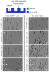
Typical fluorescence confocal micrographs show adherent cells on patterned structures. The substrate surfaces contain Ta-coated trenches and silicon oxide lines. Images of adherent cells on substrates with fixed trench widths of 1 μm are shown in (ac). Cells on patterns with fixed line widths of 1 μm are displayed on the left panel (df). Scale bars correspond to 20 μm. The DNA molecules were stained with DAPI. Phalloidin fluorescence stains were used to label actin microfilaments.
Figure 7
Figure 7
Typical scanning electron micrographs reveal adherent cells on engineered structures. Figures (ad) show comb structures consisting of a fixed trench width of 1 μm with increasing line widths of 3 μm, 5 μm, 9 μm, and 50 μm, respectively. Cells on inverted design pattern structures containing line widths of 1 μm and trench widths of 3 μm, 5 μm, 9 μm, and 50 μm are displayed in (eh), respectively. Light gray features are trenches, while dark gray areas are lines. Micrographs show cells are less likely to orient with pattern axes with trench widths. Scale bars represent 20 μm.
Figure 8
Figure 8
The figure shows the percent cell distribution of cell orientation relative to the line axes (ϕ) after incubation on substrates with various trench and line widths. Each bin corresponds to the cell population within a ± 10-degree angular range. The trench bottom and sidewalls are covered with tantalum, while the line top surface is SiO2 [25].
Figure 9
Figure 9
Plots of the percent population of cells oriented within ± 10° of the line axes for comb structures consisting of (a) a fixed line width of 1 μm and (b) a fixed trench width of 1 μm. The solid red circles represent the data collected from the monolithic Ta comb structures. Open squares denote the results of cells adhered to tantalum/SiO2 composite surfaces. Results from references [18,25] are included in these plots as a comparison. Patterns highlighted with (*) indicate a significant difference in cell alignment induced by the two types of substrates.

Similar articles

References

    1. Aubin H., Nichol J.W., Hutson C.B., Bae H., Sieminski A.L., Cropek D.M., Akhyari P., Khademhosseini A. Directed 3D cell alignment and elongation in microengineered hydrogels. Biomaterials. 2010;31:6941–6951. doi: 10.1016/j.biomaterials.2010.05.056. - DOI - PMC - PubMed
    1. Gracz A.D., Puthoff B.J., Magness S.T. Somatic Stem Cells. Humana; Totowa, NJ, USA: 2018. Cells from Murine Intestine; pp. 89–107. - DOI - PMC - PubMed
    1. Gouveia R.M., Koudouna E., Jester J., Figueiredo F., Connon C.J. Template Curvature Influences Cell Alignment to Create Improved Human Corneal Tissue Equivalents. Adv. Biosyst. 2017;1:e1700135. doi: 10.1002/adbi.201700135. - DOI - PubMed
    1. Kimura S., Tsuchiya A., Ogawa M., Ono M., Suda N., Sekimoto K., Takeo M., Tsuji T. Tissue-scale tensional homeostasis in skin regulates structure and physiological function. Commun. Biol. 2020;3:637. doi: 10.1038/s42003-020-01365-7. - DOI - PMC - PubMed
    1. Lenne P.F., Rupprecht J.F., Viasnoff V. Cell Junction Mechanics beyond the Bounds of Adhesion and Tension. Dev. Cell. 2021;56:202–212. doi: 10.1016/j.devcel.2020.12.018. - DOI - PubMed

LinkOut - more resources