. 2025 Apr 27;14(9):3029.
doi: 10.3390/jcm14093029.

Cognitive Brain Networks and Enlarged Perivascular Spaces: Implications for Symptom Severity and Support Needs in Children with Autism

Affiliations

Cognitive Brain Networks and Enlarged Perivascular Spaces: Implications for Symptom Severity and Support Needs in Children with Autism

Stefano Sotgiu et al. J Clin Med. .

Abstract

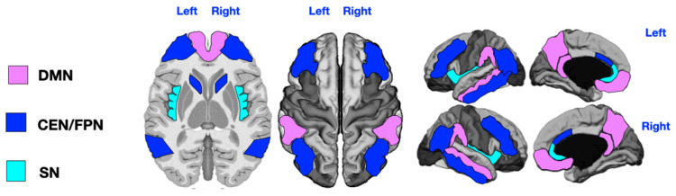
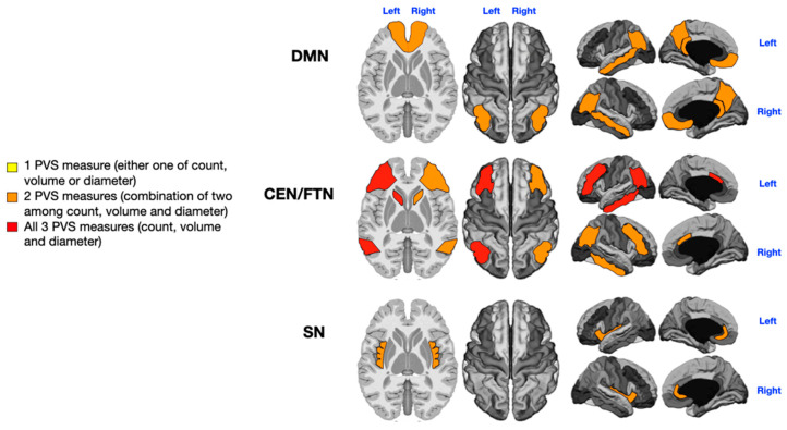
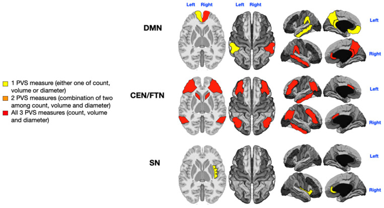
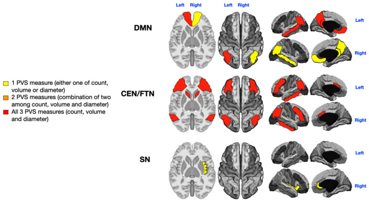
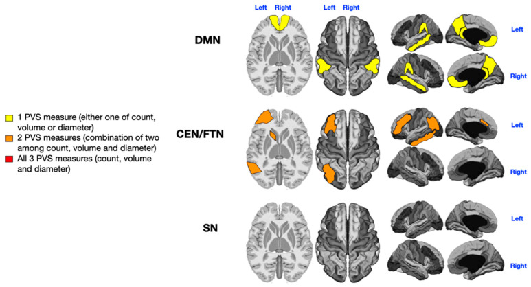
Background/Objectives: The severity of autism spectrum disorder (ASD) is clinically assessed through a comprehensive evaluation of social communication deficits, restricted interests, repetitive behaviors, and the level of support required (ranging from level 1 to level 3) according to DSM-5 criteria. Along with its varied clinical manifestations, the neuroanatomy of ASD is characterized by heterogeneous abnormalities. Notably, brain MRI of children with ASD often reveals an increased number of perivascular spaces (PVSs) compared to typically developing children. Our recent findings indicate that enlarged PVSs (ePVSs) are more common in younger male patients with severe ASD and that specific ePVS locations are significantly associated with ASD symptoms. Methods: In this study, we mapped ePVSs across key regions of three major cognitive networks-the Default Mode Network (DMN), the combined Central Executive/Frontoparietal Network (CEN/FPN), and the Salience Network (SN)-in 36 individuals with different symptom severities and rehabilitation needs due to ASD. We explored how the number, size, and location of PVSs in these networks are related to specific ASD symptoms and the overall need for rehabilitation and support. Results: Our results suggest that ePVSs in the DMN, CEN/FPN, and SN are strongly correlated with the severity of certain ASD symptoms, including verbal deficits, stereotypies, and sensory disturbances. We found a mild association between ePVSs and the level of support needed for daily living and quality of life. Conclusions: Dysfunction in cognitive networks associated with the presence of ePVSs has a significant impact on the severity of ASD symptoms. However, the need for assistance may also be influenced by other comorbid conditions and dysfunctions in smaller, overlapping brain networks.

Keywords: autism; brain functional networks; central executive network; default mode network; frontoparietal network; needs; perivascular spaces; salience network; symptoms.


Conflict of interest statement

The authors declare that there are no conflicts of interest that could prejudice the impartiality of the research reported.

Figures

Figure 1
Figure 1
Key regions of the three networks under study. DMN = Default Mode Network (purple: angular gyrus, medial orbitofrontal, middle temporal, posterior cingulate, and precuneus); CEN/FPN = Central Executive and Frontotemporal Networks (dark blue: caudal anterior cingulate, inferior parietal, inferior temporal, lateral orbitofrontal, caudate head, and rostral middle frontal); SN = Salience Network (light blue: insula and rostral anterior cingulate). The DMN key regions are angular gyrus, medial orbitofrontal cortex, middle temporal cortex, posterior cingulate, and precuneus. The CEN/FPN key regions include caudal anterior cingulate, inferior parietal cortex, inferior temporal cortex, lateral orbitofrontal cortex, caudate (head), and rostral middle frontal cortex. The SN key regions are insula and rostral anterior cingulate.
Figure 2
Figure 2
Mapping of PVS enlargement in relation to the degree of language impairment. Colored areas represent significant differences in median values (IQR) of PVS number, volume, and diameter between ASD patients with low vs. high language impairment. Areas are colored based on the extent of PVS abnormalities: yellow = single abnormality (PVS count, volume, or diameter); orange = two PVS abnormalities; red = all three PVS abnormalities. Worse language symptoms are associated with more pronounced PVS alterations, particularly in the left CEN/FPN (red).
Figure 3
Figure 3
Mapping of PVS enlargement in relation to the presence or absence of sensory disturbances. Colored areas represent significant differences in median values (IQR) of PVS number, volume, and diameter between ASD patients with low vs. high sensory disturbances. Areas are colored based on the extent of PVS abnormalities: yellow = single abnormality (PVS count, volume, or diameter); orange = two PVS abnormalities; red = all three PVS abnormalities. The presence of sensory symptoms is associated with more severe PVS alterations in both the CEN/FPN and the right DMN (red). Minimal enlargement (yellow) in the left DMN and right SN are also significantly associated with sensory issues.
Figure 4
Figure 4
Mapping of PVS enlargement in relation to the presence or absence of motor stereotypies. Colored areas represent significant differences in median values (IQR) of PVS number, volume, and diameter between ASD patients with low vs. high motor stereotypies. Areas are colored based on the extent of PVS abnormalities: yellow = single abnormality (PVS count, volume, or diameter); orange = two PVS abnormalities; red = all three PVS abnormalities. Motor stereotypies are linked to more significant PVS alterations in both the CEN/FPN and the left DMN (red). A minimal enlargement (yellow) in the right DMN and right SN are also significantly associated with motor stereotypies.
Figure 5
Figure 5
Mapping of PVS enlargement in relation to the level of rehabilitation needs. Colored areas represent significant differences in median values (IQR) of PVS number, volume, and diameter between ASD patients with low vs. high rehabilitation needs. Areas are colored based on the extent of PVS abnormalities: yellow = single abnormality (PVS count, volume, or diameter); orange = two PVS abnormalities; No areas show all three PVS abnormalities (count, volume, and diameter). Higher rehabilitation needs are associated with moderate PVS alterations, especially in the left CEN/FPN (orange). A minimal enlargement (yellow) in both DMNs is also significantly associated with rehabilitation needs.

Similar articles

References

    1. American Psychiatric Association aAPAD-TF . Diagnostic and Statistical Manual of Mental Disorders: DSM-5. American Psychiatric Association; Washington, DC, USA: 2013.
    1. Lord C., Charman T., Havdahl A., Carbone P., Anagnostou E., Boyd B., Carr T., de Vries P.J., Dissanayake C., Divan G., et al. The Lancet Commission on the future of care and clinical research in autism. Lancet. 2022;399:271–334. doi: 10.1016/S0140-6736(21)01541-5. - DOI - PubMed
    1. Zuckerman K.E., Broder-Fingert S., Sheldrick R.C. To reduce the average age of autism diagnosis, screen preschoolers in primary care. Autism. 2021;25:593–596. doi: 10.1177/1362361320968974. - DOI - PubMed
    1. Hadders-Algra M. Emerging signs of autism spectrum disorder in infancy: Putative neural substrate. Dev. Med. Child Neurol. 2022;64:1344–1350. doi: 10.1111/dmcn.15333. - DOI - PMC - PubMed
    1. McCrimmon A., Rostad K. Test Review: Autism Diagnostic Observation Schedule, Second Edition (ADOS-2) Manual (Part II): Toddler Module. J. Psychoeduc. Assess. 2013;32:88–92. doi: 10.1177/0734282913490916. - DOI

LinkOut - more resources