Review
. 2025 May 1;14(9):3145.
doi: 10.3390/jcm14093145.

Orthopedic Manifestations of Syringomyelia: A Comprehensive Review

Affiliations
Review

Orthopedic Manifestations of Syringomyelia: A Comprehensive Review

Mohamad Fadila et al. J Clin Med. .

Abstract

Background: Syringomyelia is a complex neurological disorder characterized by a fluid-filled cavity (syrinx) within the spinal cord, frequently resulting from altered cerebrospinal fluid (CSF) dynamics. While its clinical manifestations are diverse, orthopedic complications such as scoliosis, pes cavus, and Charcot arthropathy may represent early diagnostic clues yet are often under-recognized. Methods: This comprehensive review synthesizes the current literature on the pathophysiology, clinical presentation, diagnostic strategies, and management approaches of syringomyelia, with a specific emphasis on its orthopedic manifestations. Additionally, we present a detailed case of neuropathic shoulder arthropathy associated with advanced syringomyelia. Results: Orthopedic involvement in syringomyelia includes progressive spinal deformities and neurogenic joint destruction, particularly affecting the shoulder and elbow. Scoliosis is frequently observed, especially in association with Chiari malformations, and may precede neurologic diagnosis. Charcot joints result from impaired proprioception and protective sensation. The case presented illustrates the diagnostic challenges and therapeutic dilemmas in managing advanced neuro-orthopedic complications in syringomyelia. Conclusions: Syringomyelia should be considered in the differential diagnosis of atypical musculoskeletal presentations. Early recognition and multidisciplinary management are essential to prevent irreversible orthopedic sequelae. Conservative treatment remains the mainstay in stable cases, while surgery is reserved for progressive disease. Orthopedic assessment plays a pivotal role in the diagnostic pathway and long-term care of affected patients.

Keywords: Charcot joint; Chiari malformation; cerebrospinal fluid; neurogenic arthropathy; neuropathic pain; orthopedic procedures; scoliosis; spasticity; spinal cord diseases; syringomyelia.


Conflict of interest statement

The authors declare no conflicts of interest.

Figures
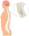
Figure 1
Figure 1
Schematic illustration of syringomyelia demonstrating the formation of a fluid-filled syrinx cavity within the spinal cord. The zoomed-in view highlights the syrinx expanding within the thoracic spinal cord, compressing adjacent neural structures and disrupting cerebrospinal fluid (CSF) dynamics—one of the primary mechanisms underlying the development of syringomyelia.
Figure 2
Figure 2
Cross-sectional schematic of the spinal cord illustrating the anatomical impact of syringomyelia. The expanding syrinx centrally disrupts the decussating fibers of the lateral spinothalamic tracts, leading to dissociated sensory loss—typically affecting pain and temperature sensation. The dorsal columns, lateral corticospinal tracts, and anterior horns may also become progressively compromised as the syrinx enlarges, resulting in motor weakness and additional sensory deficits.
Figure 3
Figure 3
Cross-sectional spinal cord diagram demonstrating the anatomical disruption caused by an expanding syrinx. The syrinx originates centrally and enlarges outward, compressing surrounding neural structures. Early involvement of the decussating spinothalamic fibers explains the characteristic dissociated sensory loss (pain and temperature), while further expansion may affect the lateral corticospinal tracts (motor pathways) and anterior horn cells (lower motor neurons), contributing to progressive motor deficits.
Figure 4
Figure 4
MRI of the thoracic spine in a patient with syringomyelia. (A) Sagittal T2-weighted image demonstrating an elongated, hyperintense intramedullary cavity consistent with a syrinx extending along the thoracic spinal cord. (B) Axial T2-weighted image showing a well-defined syrinx centrally located within the spinal cord, causing expansion of the cord parenchyma.
Figure 5
Figure 5
Radiographic and 3D imaging of a neurogenic (Charcot) shoulder joint in a patient with syringomyelia. (A) Plain radiograph showing marked destruction of the humeral head, joint subluxation, and loss of normal articular contours. (B) Three-dimensional reconstructed CT image highlighting severe bony fragmentation, deformity, and intra-articular debris consistent with advanced neuropathic arthropathy.
Figure 6
Figure 6
Suggested flowchart for the diagnostic workup of suspected syringomyelia in patients presenting with orthopedic symptoms. Multidisciplinary collaboration is recommended at each step to optimize patient outcomes.

References

    1. Leclerc A., Matveeff L., Emery E. Syringomyelia and hydromyelia: Current understanding and neurosurgical management. Rev. Neurol. 2021;177:498–507. doi: 10.1016/j.neurol.2020.07.004. - DOI - PubMed
    1. Roy A.K., Slimack N.P., Ganju A. Idiopathic syringomyelia: Retrospective case series, comprehensive review, and update on management. Neurosurg. Focus. 2011;31:E15. doi: 10.3171/2011.9.FOCUS11198. - DOI - PubMed
    1. Sabellano Z., Batino L., Navarro J.J. Atypical presentation of syringomyelia mimicking as motor neuron disease: A case report. Clin. Imag. Med. Case Rep. 2023;4:2685. doi: 10.52768/2766-7820/2685. - DOI
    1. Heiss J.D., Snyder K., Peterson M.M., Patronas N.J., Butman J.A., Smith R.K., DeVroom H.L., Sansur C.A., Eskioglu E., Kammerer W.A., et al. Pathophysiology of primary spinal syringomyelia. J. Neurosurg. Spine. 2012;17:367–380. doi: 10.3171/2012.8.SPINE111059. - DOI - PMC - PubMed
    1. Blegvad C., Grotenhuis J., Juhler M.J. Syringomyelia: A practical, clinical concept for classification. Acta Neurochir. 2014;156:2127–2138. doi: 10.1007/s00701-014-2229-z. - DOI - PubMed

LinkOut - more resources