. 2025 Dec;17(1):2505119.
doi: 10.1080/19490976.2025.2505119. Epub 2025 May 13.

Faecalibacterium prausnitzii prevents age-related heart failure by suppressing ferroptosis in cardiomyocytes through butyrate-mediated LCN2 regulation

Affiliations

Faecalibacterium prausnitzii prevents age-related heart failure by suppressing ferroptosis in cardiomyocytes through butyrate-mediated LCN2 regulation

Yun Zhang et al. Gut Microbes. 2025 Dec.

Abstract

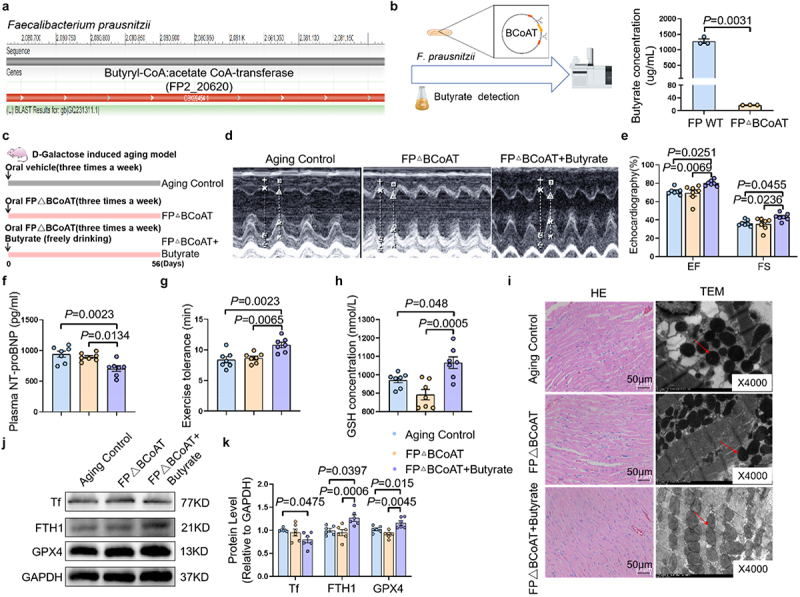
Aging is a primary driver of the escalating prevalence of heart failure (HF). Age-associated gut microbiota dysbiosis has been implicated in various age-related diseases, yet its role in age-related HF remains largely unexplored. In this study, we sought to explore the potential link between age-related gut microbiota alterations and HF in the elderly. We analyzed a publicly available single-cell sequencing dataset, which revealed markedly increased ferroptosis activity in cardiac myocytes of elderly individuals compared to their younger counterparts. Notably, treatment with the ferroptosis inhibitor, ferrostatin-1, mitigated cardiac ferroptosis and prevented cardiac dysfunction in aging rats. Furthermore, fecal microbiota transplantation from elderly HF patients significantly increased cardiac ferroptosis activity and induced cardiac dysfunction in healthy recipient rats. Integrated 16S rRNA sequencing and PCR quantification revealed a marked depletion of Faecalibacterium prausnitzii (F. prausnitzii) in elderly individuals, with a more pronounced decline in elderly patients with HF. Oral administration of F. prausnitzii or its metabolite butyrate effectively attenuated age-related HF through inhibiting ferroptosis. Additionally, gene-editing techniques were employed to generate F. prausnitzii BCoAT mutant deficient in butyrate production. Intriguingly, the protective effect was lost in the butyrate-deficient F. prausnitzii strain. Mechanistically, butyrate reduced intracellular iron accumulation and suppressed ferroptosis by downregulating LCN2 expression in senescent cardiomyocytes. Our findings highlight the critical role of aged microbiota-induced ferroptosis in HF and propose F. prausnitzii or butyrate may serve as potential targets for the prevention and treatment of age-related HF.

Keywords: F. prausnitzii; Heart failure; aging; butyrate; ferroptosis.


Conflict of interest statement

No potential conflict of interest was reported by the author(s).

Figures

Figure 1.
Figure 1.
Age-related gut microbiota increases cardiac ferroptosis and promotes heart failure. (a) UMAP plot of single-cell RNA sequencing data showing the clustering of different cell populations in heart tissue of young and elderly individuals. (b) KEGG pathway enrichment analysis showed the pathways significantly associated with gene expression changes in ventricular cardiac myocytes. (c) Representative image of Perls’ blue staining in the left ventricle of young and aging rats. (d) Schematic illustration of the experimental design for ferroptosis inhibitor intervention in aging rats. 18 months old rats were randomly subjected to receive vehicle or ferroptosis inhibitor (ferrostatin-1, 0.8 mg/kg, intraperitoneal injection), once a week for 12 weeks. (e) Representative M-mode images of left ventricular wall motion in the hearts of rats from aging+Vehicle group and aging+Fer-1 group. (f) Quantification of cardiac function parameters, cardiac ejection fraction (EF) and cardiac fraction shortening (FS) of rats from aging+Vehicle group and aging+Fer-1 group (n = 6 per group). (g) Quantitative analysis of plasma NT-proBNP levels in rats from aging+Vehicle group and aging+Fer-1 group (n = 6 per group). (h) The exercise tolerance in rats (n = 6 per group). (i) Representative images of HE staining of the left ventricle of hearts in rats. (j) Schematic illustration of the experimental design for microbial transplantation experiments. The feceal samples were collected from young and aging rats for microbial transplantation experiments. Before microbial transplantation, rats were treated with antibiotics for 1 week to deplete gut microbiota. Afterwards, microbiota-depleted rats received microbiota from young rats, or aging rats through gavage, once a day for 12 weeks. (k) Representative M-mode images of left ventricular wall motion in the hearts of rats. (l) The statistical data of cardiac ejection fraction (EF) and fraction shortening (FS) of rats (n = 8 per group). (m) Quantitative analysis of plasma NT-proBNP levels in rats (n = 8 per group). (n) The exercise tolerance in rats (n = 6 per group). (o) Quantitative analysis of GSH levels in rats (n = 8 per group). (p) Representative images of HE staining of the left ventricle, and representative transmission electron microscopy images of mitochondria in the left ventricle of rats.
Figure 2.
Figure 2.
F. prausnitzii inhibits cardiac ferroptosis and prevents the development of age-related heart failure. (a) The PCoA showed that the gut taxonomic composition was significantly different between young (n = 250) and elderly individuals (n = 98). (b) The alpha diversity of microbial community in young and elderly individuals. (c) Secondary analysis of previously published 16S rRNA sequencing data showed the gut microbiota composition between young and elderly individuals (DRA004160-). (d) Schematic diagram of the experiment: fecal samples were collected from healthy elderly individuals and age- and sex-matched elderly heart failure patients to analyze the abundance of F. prausnitzii. Clinical information was also collected. (e) The relative abundance of F. prausnitzii in elderly healthy individuals and patients with elderly HF (n = 40 per group). (f) ROC curve analysis of F. prausnitzii abundance for the diagnosis of heart failure, showing an AUC of 0.703 (P = 0.002, 95% CI: 0.589–0.816). (g) Correlation analysis between F. prausnitzii abundance and ejection fraction (EF%), showing a positive correlation (R = 0.096, P = 0.005). (h) Schematic illustration of the experimental design. We established an aging rat model with D-galactose (150 mg/kg/day) via subcutaneous injection. The rats were randomly divided into a control group, a D-galactose-induced aging rats group, and a D-galactose-induced aging rats supplemented with F. prausnitzii group. (i) Representative M-mode images of left ventricular wall motion in the hearts, and the statistical data of cardiac ejection fraction (EF) and fraction shortening (FS) of rats (n = 8 per group). (j) Quantitative analysis of plasma NT-proBNP levels in rats (n = 8 per group). (k) The exercise tolerance in rats (n = 8 per group). (l) The levels of GSH in rats (n = 8 per group). (m) Representative images of HE staining, and representative transmission electron microscopy images of mitochondria in the hearts in rats. (n) Representative bands and quantification of expressions of Tf, FTH1 and GPX4 in the hearts of rats (n = 6 per group).
Figure 3.
Figure 3.
F. prausnitzii-derived butyrate alleviates ferroptosis and prevents age-related heart failure. (a) Cellular experiment schematic: H2O2 was used to establish a senescent cell model, and subsequently exposed to live F. prausnitzii, inactivated F. prausnitzii, and F. prausnitzii supernatant. (b) The cell viability of cardiomyocytes (n = 6 per group). (c) Representative images of FerroOrange detection in cardiomyocytes. (d) Butyrate detection by mass spectrometry: media (control) and F. prausnitzii supernatant (FP SUP) were collected for analysis to measure butyrate levels. (e) The concentration of butyrate in the media and F. prausnitzii supernatant (n = 7 per group). (f) The cell viability of cardiomyocytes in control group, H2O2 group, and H2O2 + butyrate group (n = 7 per group). (g) Representative image of FerroOrange detection in cardiomyocytes. (h) Schematic illustration of the experimental design. We established an aging rat model with D-galactose (150 mg/kg/day) via subcutaneous injection. The rats were randomly divided into two groups, received water containing butyrate sodium (1% w/w) for 8 weeks (Aging+butyrate group), another group received ordinary water (Aging+veh group). (i) Representative images of M-mode echocardiograms of hearts, and the statistical data of cardiac ejection fraction (EF) and cardiac fraction shortening (FS) of rats (n = 8 per group). (j) The plasma levels of NT-proBNP in rats (n = 8 per group). (k) The exercise tolerance in rats (n = 8 per group). (l) The levels of GSH in rats (n = 8 per group). (m) Representative images of HE staining, and transmission electron microscopy images of mitochondria in the heart of rats.
Figure 4.
Figure 4.
F.Prausnitzii prevents age-related heart failure through butyrate. (a) The genome of F.Prausnitzii L2–6 contained FP2_20620 encoding butyryl-CoA:acetate CoA-transferase. (b) Quantification of the concentration of butyrate in supernatant of F.P and F.PΔBCoAT assessed by mass spectrometry (n = 4 per group). (c) Schematic illustration of the experimental design. We established an aging rat model with D-galactose (150 mg/kg/day) via subcutaneous injection. The rats were randomly divided into three groups, one group received F.PΔBCoAT gavage for 8 weeks (F.PΔBCoAT group), another group received F.PΔBCoAT gavage combined with water containing butyrate sodium (1% w/w) for 8 weeks (F.PΔBCoAT+Butyrate group), the aging control group received ordinary water. (d) Representative images of M-mode echocardiograms of hearts in rats. (e) The statistical data of cardiac ejection fraction (EF) and cardiac fraction shortening (FS) of rats (n = 7 per group). (f) The plasma levels of NT-proBNP in rats (n = 7 per group). (g) The exercise tolerance in rats (n = 7 per group). (h) The levels of GSH in rats (n = 7 per group). (i) Representative images of HE staining, and representative transmission electron microscopy images of mitochondria in the left ventricle of hearts in rats. The red arrow points to the mitochondria. (j) Representative bands showing the expressions of tf, FTH1 and GPX4 in the hearts of rats. (k) Quantification of the expressions of tf, FTH1 and GPX4 in the hearts of rats (n = 6 per group).
Figure 5.
Figure 5.
Butyrate inhibits ferroptosis by modulating LCN2 in cardiomyocytes. (a) Transcriptomics was performed to detect the gene expression changes. The volcano plot showed diferentially expressed genes between Aging+Veh group and Aging+Butyrate group. n = 4 biological replicates/group. (b) The statistical data shows the 213 up-regulated genes, 434 down-regulated genes between the two groups. (c) Gene ontology (GO) analysis for upregulated and downregulated genes in the RNA-seq data. (d) Representative ROS staining images and fluorescence intensity in the left ventricle of heart in rats. (e) Representative ROS staining images of cardiomyocytes. (f) Analysis of significantly differentially expressed genes in the oxidative stress pathway in the left ventricle of hearts of rats from Aging+Veh group and Aging+Butyrate group. (g) Cardiomyocytes were transfected with plasmids to silence LCN2. After 24 h, they were treated with H2O2 for 6 h. Following a change of culture media, the cells were further incubated for 42 h before conducting assays for relevant markers. (h) The viability of cardiomyocytes (n = 6 per group). (i) Representative image of FerroOrange detection in cardiomyocytes. (j) Cardiomyocytes were first transfected with plasmids for LCN2 overexpression. After 24 h, they were treated with H2O2 for 6 h. Following a change of culture media, the cells in H2O2+butyrate group and H2O2+butyrate+OE-LCN2 group were treated with butyrate for 42 h before conducting assays for relevant markers. The viability of cardiomyocytes (n = 6 per group). (k) Representative image of FerroOrange detection in cardiomyocytes.
Figure 6.
Figure 6.
Faecalibacterium prausnitzii prevents age-related heart failure through inhibition of ferroptosis by butyrate. Aged microbiota dysbiosis heightens ferroptosis and increases susceptibility to HF in rats. Oral supplementation of F. prausnitzii mitigates ferroptosis and confers protection against age-related HF through butyrate by downregulating LCN2 in senescent cardiomyocyte.

References

    1. McDonagh TA, Metra M, Adamo M, Gardner RS, Baumbach A, Böhm M, Burri H, Butler J, Čelutkienė J, Chioncel O, et al. 2021 ESC guidelines for the diagnosis and treatment of acute and chronic heart failure. Eur Heart J. 2021;42(36):3599–20. doi: 10.1093/eurheartj/ehab368. - DOI - PubMed
    1. Roger VL. Epidemiology of heart failure: a contemporary perspective. Circ Res. 2021;128(10):1421–1434. doi: 10.1161/CIRCRESAHA.121.318172. - DOI - PubMed
    1. Savarese G, Becher PM, Lund LH, Seferovic P, Rosano G, Coats A. Global burden of heart failure: a comprehensive and updated review of epidemiology. Cardiovasc Res. 2023;118(17):3272–3287. doi: 10.1093/cvr/cvac013. - DOI - PubMed
    1. Luo Y, Zhang Y, Han X, Yuan Y, Zhou Y, Gao Y, Yu H, Zhang J, Shi Y, Duan Y, et al. Akkermansia muciniphila prevents cold-related atrial fibrillation in rats by modulation of TMAO induced cardiac pyroptosis. EBioMedicine. 2022;82:104087. doi: 10.1016/j.ebiom.2022.104087. - DOI - PMC - PubMed
    1. Zhou Q, Deng J, Pan X, Meng D, Zhu Y, Bai Y, Shi C, Duan Y, Wang T, Li X, et al. Gut microbiome mediates the protective effects of exercise after myocardial infarction. Microbiome. 2022;10(1):82. doi: 10.1186/s40168-022-01271-6. - DOI - PMC - PubMed

MeSH terms