. 2025 Jun;14(15):e2500107.
doi: 10.1002/adhm.202500107. Epub 2025 May 13.

Engineered Chitosan-Derived Nanocarrier for Efficient siRNA Delivery to Peripheral and Central Neurons

Affiliations

Engineered Chitosan-Derived Nanocarrier for Efficient siRNA Delivery to Peripheral and Central Neurons

Ana P Spencer et al. Adv Healthc Mater. 2025 Jun.

Abstract

Gene therapy using small interfering RNA (siRNA) holds promise for treating neurological disorders by silencing specific genes, like the phosphatase and tensin homolog (PTEN) gene, which restricts axonal growth. Effective siRNA delivery to neurons, however, poses challenges due to premature nucleic acid degradation and unspecific delivery. Chitosan-based delivery systems, noted for their biocompatibility, face limitations such as low transfection efficiency and lack of neurotropism. Building on the previous successes with neuron-targeted DNA delivery using chitosan, a novel approach for siRNA delivery aimed at PTEN downregulation is proposed. This involves using thiolated trimethyl chitosan (TMCSH)-based siRNA nanoparticles functionalized with the neurotropic C-terminal fragment of the tetanus neurotoxin heavy chain (HC) for efficient delivery to peripheral and central neurons. These polyplexes demonstrate suitable physicochemical properties, biocompatibility, and no adverse effects on neuronal electrophysiology. Diverse neuronal models, including 3D ex vivo cultures and microfluidics, confirm the polyplexes' efficiency and neurospecificity. HC-functionalization significantly enhances neuronal binding, and live cell imaging reveals fivefold faster retrograde transport along axons. Furthermore, siRNA delivery targeting PTEN promoted axonal outgrowth in embryonic cortical neurons. In conclusion, the proposed polyplexes represent a promising platform for neuronal siRNA delivery, offering potential for clinical translation and therapeutic applications.

Keywords: PTEN; chitosan; gene therapy; microfluidics; siRNA.


Conflict of interest statement

The authors declare no conflict of interest.

Figures
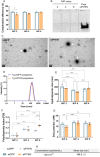
Figure 1
Figure 1
Physicochemical characterization of targeted (Tg) polyplexes prepared with siGFP or siPTEN as a function of N/P ratio. All NPs were prepared in phosphate‐buffered saline (PBS) 1×, pH 7.4. A) SYBR Gold exclusion assay (room temperature), B) polyacrylamide gel electrophoresis (PAGE), C) transmission electron microscopy (TEM) images of Tg N/P 4 polyplexes prepared with siGFP (on the left) or with siPTEN (on the right). White arrows indicate polyplexes. Scale bar: 100 nm. D) Representative dynamic light scattering (DLS) curves of Tg polyplexes carrying siGFP or siPTEN, E) size distribution (DLS), F) polydispersity index (PDI, DLS), G) surface charge (Electrophoretic Light Scattering), H) concentration and mean size of Tg N/P 4 polyplexes in solution (nanoparticle tracking analysis, NTA). Results are shown as mean ± SD (standard deviation) of three independent experiments (n = 3) with A) two, D–G) one sample, and three measurements per sample or H) one sample, per experiment. For statistical analysis, a two‐way ANOVA test was used. Significant differences: *p < 0.05, **p < 0.01, and ****p ≤ 0.0001. Results of the physicochemical characterization of non‐targeted (nTg) polyplexes can be found in Table S2 (Supporting Information).
Figure 2
Figure 2
Cellular binding, internalization, and downregulation capacity of polyplexes. A) ND7/23, HT22, and NIH 3T3 cells were incubated with targeted (Tg) polyplexes (N/P 2, 4, and 6) carrying Cy5‐labeled siRNAmi for 8 h (final siRNAmi concentration 100 nm). Characterization by flow cytometry in terms of Cy5 positive cells (in %), B) a similar cellular association assay was done but including a pre‐incubation with HC (final concentration 1 nm) for 15 min at 4  °C, C) representative images of entire embryonic DRG explant untreated (on the left) or incubated, for 24 h, with Cy5‐siRNAmi (on the center) and with Tg polyplexes carrying Cy5‐siRNAmi (final concentration 100 nm, on the right). Staining: βIII‐tubulin (yellow), nuclei with Hoechst 33 342 (gray), and Cy5‐siRNAmi (pink). White arrows indicate Cy5 signal associated with the polyplexes in the DRG explant body Scale bar: 200 µm, D) Cy5 fluorescence intensity quantification in the DRG explant body. E) Downregulation of GFP expression measured in motor neurons after 96‐hour incubation with N/P 4 polyplexes. Results are represented as mean ± SD of three independent experiments (n = 3), with two replicates per experiment. For statistical analysis, a two‐way ANOVA test was used. Significant differences: *p < 0.05, and ****p ≤ 0.0001.
Figure 3
Figure 3
Schematic representation of the microfluidic chips design and pictures of the poly(dimethylsiloxane) (PDMS) microfluidic devices employed in this study. A) Two‐compartment microfluidics with 450 µm microchannels. Compartment 1 refers to the cell soma compartment, while compartment 2 is the compartment for neuronal axon terminals, B) three‐compartment microfluidics with two sets of 600 µm microchannels. Compartments 1 and 3 refer to the compartments for neuronal axon terminals, and compartment 2 is the cell soma compartment of the neurons.
Figure 4
Figure 4
Polyplexes interaction with neurons, under flow conditions. Motor or cortical neurons were seeded in two‐compartment microfluidics and incubated overnight underflow with Tg and nTg N/P 4 polyplexes carrying Cy5‐labeled siRNAmi (added in the axonal compartment, final concentration 100 nm). A) Schematic representation of the setup, B) side‐view illustration of nanoparticle incubation underflow in microfluidic,(C) representative image of cortical neurons incubated with Tg N/P 4 polyplexes carrying Cy5‐siRNAmi. Staining: βIII‐tubulin (yellow), nuclei with Hoechst 33 342 (cyan), and Cy5‐siRNAmi (pink). White arrows indicate Cy5 signal associated with the polyplexes. Scale bar: 25 µm, D) the number of puncta inside neurons (cell soma compartment) was quantified. Results are represented as mean ± SD of three independent experiments (n = 3), with one replicate per experiment. For statistical analysis, a two‐way ANOVA test was used. Significant differences: **p < 0.01.
Figure 5
Figure 5
Retrograde axonal transport characteristics in motor and cortical neurons. The probability density function of the instantaneous velocity of nTg and Tg N/P 4 polyplexes‐loaded vesicles in A) motor neurons and B) cortical neurons, defined as the speed (µm s−1) of movement between consecutive images within a sequence, C) average velocity (µm s−1) of retrograde axonal transport of NPs‐loaded vesicles, D) total run length and E) total pause periods. Results are represented as mean ± SD of three independent experiments (n = 3), with one replicate per experiment. One‐way ANOVA tests were used for statistical analysis. Significant differences: *p < 0.05, **p < 0.01, and ****p ≤ 0.0001.
Figure 6
Figure 6
The cellular interaction kinetics of polyplexes in microfluidic‐based cultures. A) Representative images from confocal microscopy of Tg N/P 4 polyplexes carrying Cy5‐siRNAmi. Staining: Nuclei with Hoechst 33 342 (cyan), βIII tubulin (yellow), and Cy5‐siRNAmi polyplexes (pink). Through image analysis, the fluorescence intensity in the cell bodies of B) motor neurons and C) cortical neurons was quantified. Results are represented as the mean ± SD of three independent experiments (n = 3), with at least six replicates per experiment. For statistical analysis, a one‐way ANOVA test was used. Significant differences: *p < 0.05 and **p < 0.01.
Figure 7
Figure 7
Nanoparticle biocompatible profile evaluation in microfluidic‐based primary motor and cortical neurons. Electrical activity evaluation through the total number of spikes of A) motor neurons and B) cortical neurons cultured in two‐compartment microfluidic. Results are represented as the mean ± SD of three independent experiments (n = 3), with three replicates per experiment. For statistical analysis, a two‐way ANOVA test was used. Significant differences: ****p ≤ 0.0001.
Figure 8
Figure 8
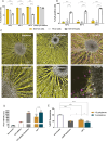
The biological effect of NPs formulations in primary cortical neurons. A) Representative mosaic image used to quantify axonal outgrowth, in three‐compartment microfluidic devices, after treatment with targeted N/P 4 polyplexes carrying siPTEN. Staining: Neurofilament H (in yellow). B) Average axon length quantified in neurons growing in three‐compartment microfluidic cultures, C) number of axons per microchannel, and D) number of intersections of the axons in linear grids spaced 50 µm apart. Results are represented as the mean ± SD of three independent experiments (n = 3), with three replicates per experiment. For statistical analysis, a two‐way ANOVA test was used. Significant differences: *p < 0.05, **p < 0.01, *** p < 0.001 and ****p ≤ 0.0001. In C) and D), significant differences are observed compared to untreated cells.

References

    1. Sudhakar V., Richardson R. M., Neurotherapeutics 2019, 16, 166. - PMC - PubMed
    1. Pena S. A., Iyengar R., Eshraghi R. S., Bencie N., Mittal J., Aljohani A., Mittal R., Eshraghi A. A., J. Drug Targeting 2020, 28, 111. - PubMed
    1. Luo M., Lee L. K. C., Peng B., Choi C. H. J., Tong W. Y., Voelcker N. H., Adv. Sci. 2022, 9, 2201740. - PMC - PubMed
    1. Park K. K., Liu K., Hu Y., Smith P. D., Wang C., Cai B., Xu B., Connolly L., Kramvis I., Sahin M., He Z., Science 2008, 322, 963. - PMC - PubMed
    1. Ning K., Drepper C., Valori C. F., Ahsan M., Wyles M., Higginbottom A., Herrmann T., Shaw P., Azzouz M., Sendtner M., Hum. Mol. Genet. 2010, 19, 3159. - PubMed