. 2024 Nov 4;49(2):148-161.
doi: 10.55730/1300-0152.2733. eCollection 2025.

Progression of electrophysiological impairments in diabetic cardiomyopathy and intervention using Enicostemma axillare

Affiliations

Progression of electrophysiological impairments in diabetic cardiomyopathy and intervention using Enicostemma axillare

Victor Arokia Doss et al. Turk J Biol. .

Abstract

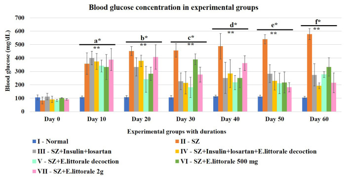
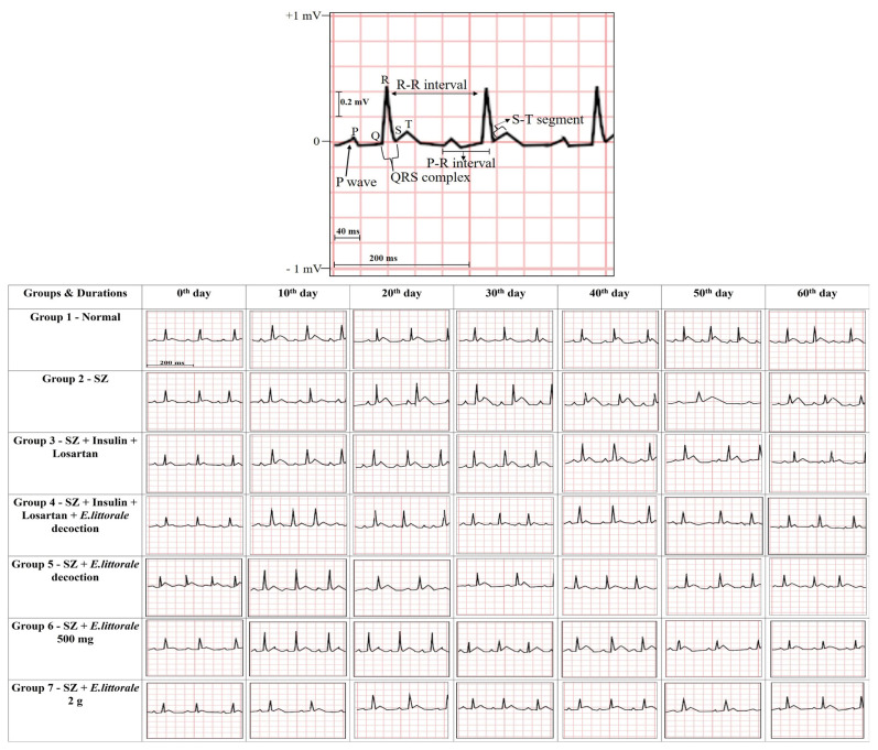
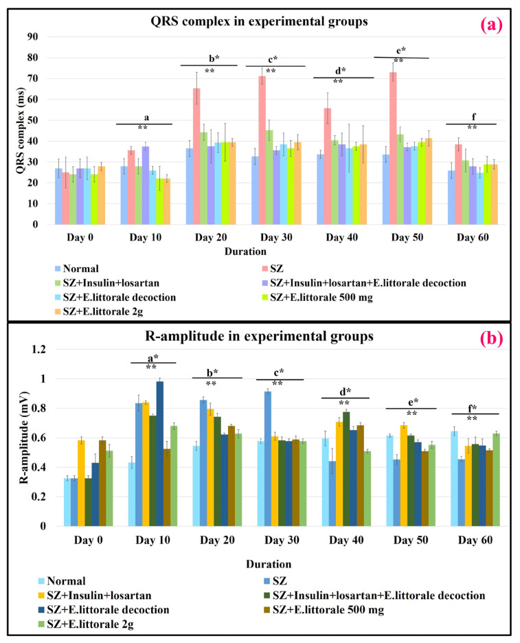
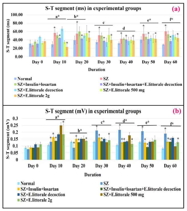
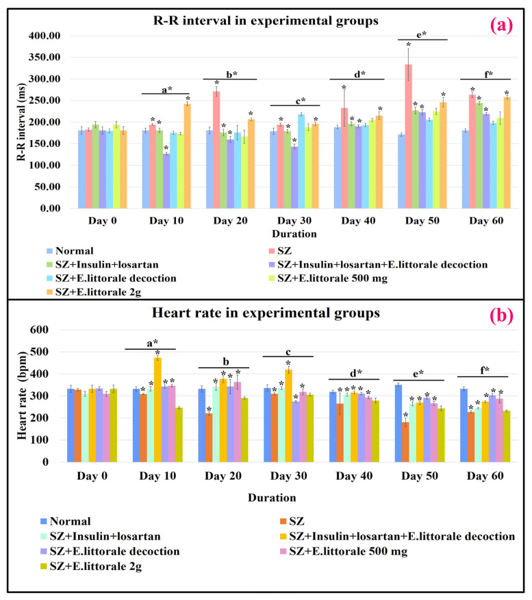
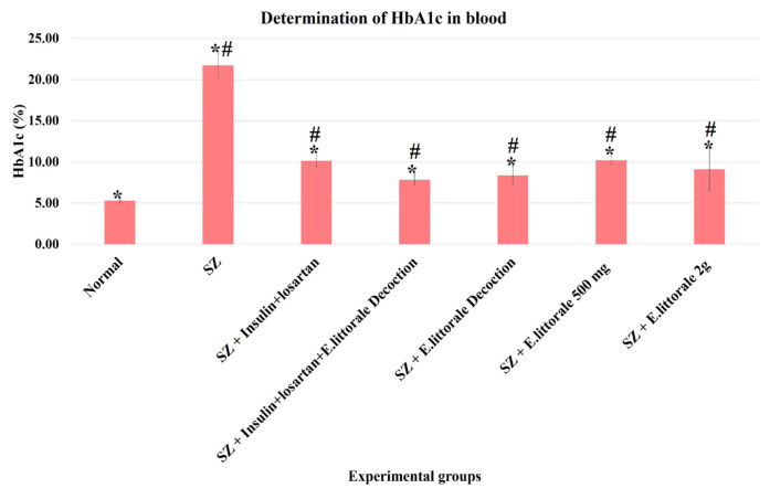
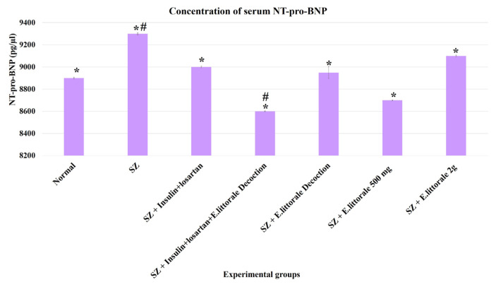
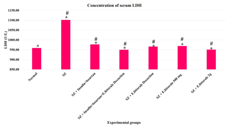
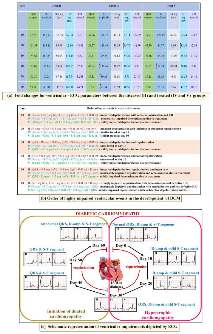
Earlier studies widely reported advanced ventricular impairments only at the later stages of diabetic cardiomyopathy (DCM). The present study aimed to understand the duration-based early to late stages of cardiac electrophysiological impairments using electrocardiography (ECG) and intervention using Enicostemma axillare subsp. littorale. The experimental rats were streptozotocin (SZ)-induced diabetic rats that were administered E. axillare formulations as follows: (1) normal; (2) SZ - 40 mg/kg, single, i.p.); (3) SZ + insulin (2 U/day) + losartan (10 mg/kg); (4) SZ + insulin + losartan + E. axillare decoction (2 mL/day); (5) SZ + E. axillare decoction; (6) SZ + E. axillare (low dose-500 mg/kg), and (7) SZ + E. axillare (high dose-2 g/kg). Steady hyperglycemia was witnessed until day 5 followed by various elevated patterns until day 60 in the SZ group that was potentially treated with E. axillare formulations from day 20. ECG (lead II) revealed early significantly impaired ventricular events, namely widened QRS complex, elevated R-amplitude, and prolonged R-R interval from day 10 that were regulated using E. axillare decoction. Pearson's correlation analysis revealed a strong relationship between basal blood glucose (day 0) and impaired ECG parameters. This duration-based study therefore illustrated the progression of glycemic shifts, ventricular impairments, and their correlation besides establishing the interventional potential of E. axillare subsp. littorale towards these impairments associated with DCM. These glycemic-ECG impairments were substantiated by the increased blood HbA1c, serum NT-pro BNP, and LDH levels that were ameliorated by E. axillare subsp. littorale decoction. It was concluded that E. axillare subsp. littorale can restore early ventricular depolarization impairments, marking it as the reversible hypertrophic cardiomyopathic stage towards intervention in DCM.

Keywords: Blood glucose; cardiac hypertrophy; diabetes; diabetic cardiomyopathy; electrocardiography.


Conflict of interest statement

Conflicts of interest: The authors declare no conflict of interest. The funders had no role in the design of the study; in the collection, analyses, or interpretation of data; in the writing of the manuscript, or in the decision to publish the results.

Figures

Figure 1
Figure 1
Chronic hyperglycemia at various durations. SZ rats (group II) exhibited more significant hyperglycemia than normal rats (group I). All the treated groups (III to VII) especially with E. axillare subsp. littorale decoction significantly reduced this elevation preferably from day 20 onwards in group VI. Two-way ANOVA for early durations of blood glucose showed significant relationship between experimental groups and the durations [F (78,294) = 9.805, p = 3.447E-48] at every 10 day-interval [F (36,147) = 6.514, p = 1.1153E-16]. This indicated that the efficacy of the experimental conditions varies in different durations based upon the respective potentials towards induction or treatment of hyperglycemia.
Figure 2
Figure 2
Graphical representation of ECG recordings at various durations. P wave: atrial depolarization; P–R interval: atrioventricular propagation of impulse; QRS complex and R-amplitude: ventricular depolarization; ST segment: ventricular repolarization; R–R interval: duration between two heart beats/heart rate (HR).
Figure 3
Figure 3
ECG parameters of ventricular depolarization. Delayed communication between ventricular cardiomyocytes was significantly observed by widened QRS and elevated R-amplitude from day 20 in diseased (SZ-group II) rats that were regulated by E. axillare subsp. littorale formulations. Two-way ANOVA for the QRS complex [F (36,147) = 6.092, p = 1.43E-15] and R-amplitude [F (36,147) = 61.581, p = 7.6532E-72] showed significant interactions between experimental conditions (groups) and durations that can influence the ventricular events (QRS complex and R-amplitude) together. This indicated that the experimental conditions and different durations are associated with changes in QRS complex and R-amplitude.
Figure 4
Figure 4
ECG parameters for ischemic conditions. The disease group (SZ-group II) exhibited a significantly elevated ST segment (ms and mV) indicating the presence of ischemia and impaired ventricular events that were significantly reduced by coadministration of E. axillare subsp. littorale decoction with insulin and losartan (group IV). Relationships between experimental groups and durations were significant for ST segment (ms) [F (36,147) = 2.638, p = 0.000024] and ST segment (mV) [F (36,147) = 8.436, p = 2.4215E-21]. This indicated that the experimental conditions and durations can influence the ST segment.
Figure 5
Figure 5
ECG parameters of heart rhythm. The disease group (SZ-group II) was observed with prolonged R–R interval embarking reduced HR (bradycardia) that were extended until day 60 and were under control in the treatment groups. Statistically significant interactions between experimental groups and durations were seen in R–R interval (ms) [F (36,147) = 8.438, p = 2.4215E-21] and heart rate [F (36,147) = 25.565, p = 4.5217E-47]. This shows that both experimental groups and durations have significant effects upon the changes in R–R interval and HR.
Figure 6
Figure 6
Determination of blood HbA1c in the experimental groups. A significantly increased HbA1c level was observed in the diseased group (SZ-group II) when compared to the normal group (group I). With comparison to the reference drugs (group III) the group coadministered E. axillare decoction and reference drugs (group IV) was highly efficient with reduced HbA1c levels. This was followed by the groups administered E. axillare decoction (group V) and low dosage extract powder (group VI).
Figure 7
Figure 7
Determination of serum NT-pro BNP in the experimental groups. Significantly increased serum NT-pro BNP level was observed in the diseased group (SZ-group II) when compared to the normal group (group I). With comparison to the reference drugs (group III) the efficacy of the plant formulations in reducing NT-pro BNP levels to near normal levels was observed to be high in the group administered E. axillare decoction (group V).
Figure 8
Figure 8
Determination of serum LDH in the experimental groups. Significantly increased serum LDH level was observed in the diseased group (SZ-group II) when compared to the normal group (group I). With comparison to the reference drugs (group III) the efficacy of the plant formulations in regulating LDH levels to near normal levels was observed to be highly potent in the groups treated with E. axillare decoction (groups IV and V) followed by the high dosage extract powder (group VII).
Figure 9
Figure 9
Overview of ECG parameters in DCM and the effect of E. axillare subsp. littorale. In the diseased group (SZ-group II) impaired ventricular depolarization (R-amplitude) was witnessed during the early days until day 30 embarking on hypertrophic state. Next, it was accompanied with widened QRS complex and elevated ST segment towards day 60, indicating dilated ventricles. Direct and coadministration of E. axillare decoction (groups IV and V) displayed a profound interventional effect upon these early ventricular hypertrophic impairments, thus preventing the progression of DCM.

Similar articles

References

    1. Abukhalil MH, Althunibat OY, Aladaileh SH, Al-Amarat W, Obeidat HM, et al. Galangin attenuates diabetic cardiomyopathy through modulating oxidative stress, inflammation and apoptosis in rats. Biomedicine & pharmacotherapy = Biomedecine & Pharmacotherapie. 2021;138:111410. doi: 10.1016/j.biopha.2021.111410. - DOI - PubMed
    1. Al-Fahham AA. Development of New LSD Formula when Numbers of Observations Are Unequal. Open Journal of Statistics. 2018;8:258–263. doi: 10.4236/ojs.2018.82016. - DOI
    1. Ananthakrishnan M, Doss VA. Effect of 50% hydro-ethanolic leaf extracts of Ruellia tuberosa L. and Dipteracanthus patulus(jacq.) on lipid profile in alloxan induced diabetic rats. International Journal of Preventive Medicine. 2013;4(7):744–747. - PMC - PubMed
    1. Aydin S, Ugur K, Aydin S, Sahin İ, Yardim M. Biomarkers in acute myocardial infarction: current perspectives. Vascular Health and Risk Management. 2019;15:1–10. doi: 10.2147/VHRM.S166157. - DOI - PMC - PubMed
    1. Badole LS, Chaudhari SM, Jangam GB, Kandhare DA, Bodhankar LS. Cardioprotective activity of Pongamia pinnata in streptozotocin – nicotinamide induced diabetic rats. BioMed Research International. 2015;403291:1–8. doi: 10.1155/2015/403291. - DOI - PMC - PubMed

LinkOut - more resources