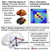
Altered functional connectivity strength between structurally and functionally affected brain regions in visual snow syndrome
- PMID: 40365132
- PMCID: PMC12069226
- DOI: 10.1093/braincomms/fcaf171
Altered functional connectivity strength between structurally and functionally affected brain regions in visual snow syndrome
Abstract
Visual snow syndrome (VSS) is a neurological disorder that is predominantly characterized by persistent, dynamic visual disturbances, experienced across the entire visual field. Earlier research highlighted the significance of distinct brain regions, exhibiting alterations in both anatomical structure and functional characteristics. To further investigate the functional role of these regions, we examined the resting-state connectivity between these areas in individuals with VSS and the relation with VSS symptoms and oculomotor measures of visual processing. Forty patients with VSS (53% females; age = 33.2 ± 10.1 years; 22 with migraine) and 60 healthy controls (58% females; age = 32.0 ± 9.2 years) were scanned using 7 Tesla MRI system. High spatial and temporal resting-state (RS) functional (TR = 800 ms, 1.6 mm isotropic) and anatomical (MP2RAGE, 0.75 mm isotropic) images were acquired. Resting-state data were pre-processed (motion correction, temporal filtering and spatial smoothing), functional connectivity was calculated between regions of interest and compared between groups. Significant metrics were compared with VSS patients with and without migraine and correlated with oculomotor measures (prosaccade and anti-saccade latencies), number of VSS symptoms, self-rated VSS intensity and perceived disruptiveness. Compared to healthy controls, VSS patients demonstrated significantly higher connectivity between the supramarginal gyrus and lateral occipital cortex (P = 0.016) and fusiform (P = 0.007), lower connectivity between the supramarginal gyrus and pallidum (P = 0.032), as well as between the parahippocampal gyrus and lateral occipital cortex (P = 0.007), which related to higher perceived disruptiveness (P = 0.002, r = -0.489). No differences were found between VSS with and without migraine. This study revealed altered functional connectivity strength in individuals with VSS, suggesting stronger connectivity between cortical areas, particularly centred around the supramarginal gyrus, and disconnections with deep grey matter and temporal cortices, which associated with perceived disruptiveness of VSS.
Keywords: 7T; fMRI; functional connectivity; resting-state MRI; visual snow syndrome.
© The Author(s) 2025. Published by Oxford University Press on behalf of the Guarantors of Brain.
Conflict of interest statement
The authors report no competing interests.
Figures


Similar articles
-
Brain network dynamics in people with visual snow syndrome.Hum Brain Mapp. 2023 Apr 1;44(5):1868-1875. doi: 10.1002/hbm.26176. Epub 2022 Dec 8. Hum Brain Mapp. 2023. PMID: 36478470 Free PMC article.
-
Microstructure in patients with visual snow syndrome: an ultra-high field morphological and quantitative MRI study.Brain Commun. 2022 Jun 23;4(4):fcac164. doi: 10.1093/braincomms/fcac164. eCollection 2022. Brain Commun. 2022. PMID: 35974797 Free PMC article.
-
Disrupted connectivity within visual, attentional and salience networks in the visual snow syndrome.Hum Brain Mapp. 2021 May;42(7):2032-2044. doi: 10.1002/hbm.25343. Epub 2021 Jan 15. Hum Brain Mapp. 2021. PMID: 33448525 Free PMC article.
-
Visual Snow Syndrome as a Network Disorder: A Systematic Review.Front Neurol. 2021 Oct 4;12:724072. doi: 10.3389/fneur.2021.724072. eCollection 2021. Front Neurol. 2021. PMID: 34671311 Free PMC article.
-
Visual Snow: Visual Misperception.J Neuroophthalmol. 2018 Dec;38(4):514-521. doi: 10.1097/WNO.0000000000000702. J Neuroophthalmol. 2018. PMID: 30095537 Review.
References
-
- Schankin CJ, Maniyar FH, Digre KB, Goadsby PJ. Visual snow”—A disorder distinct from persistent migraine aura. Brain. 2014;137(5):1419–1428. - PubMed
-
- Traber GL, Piccirelli M, Michels L. Visual snow syndrome: A review on diagnosis, pathophysiology, and treatment. Curr Opin Neurol. 2020;33(1):74–78. - PubMed
LinkOut - more resources
Full Text Sources
