

Not every nasal block is allergic rhinitis: a case report of sinonasal malignant melanoma
- PMID: 40365267
- PMCID: PMC12069232
- DOI: 10.1093/jscr/rjaf295
Not every nasal block is allergic rhinitis: a case report of sinonasal malignant melanoma
Abstract
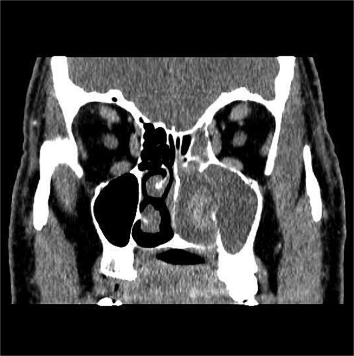
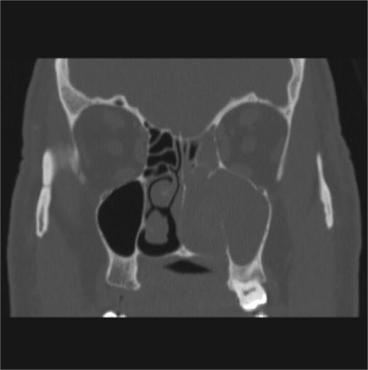
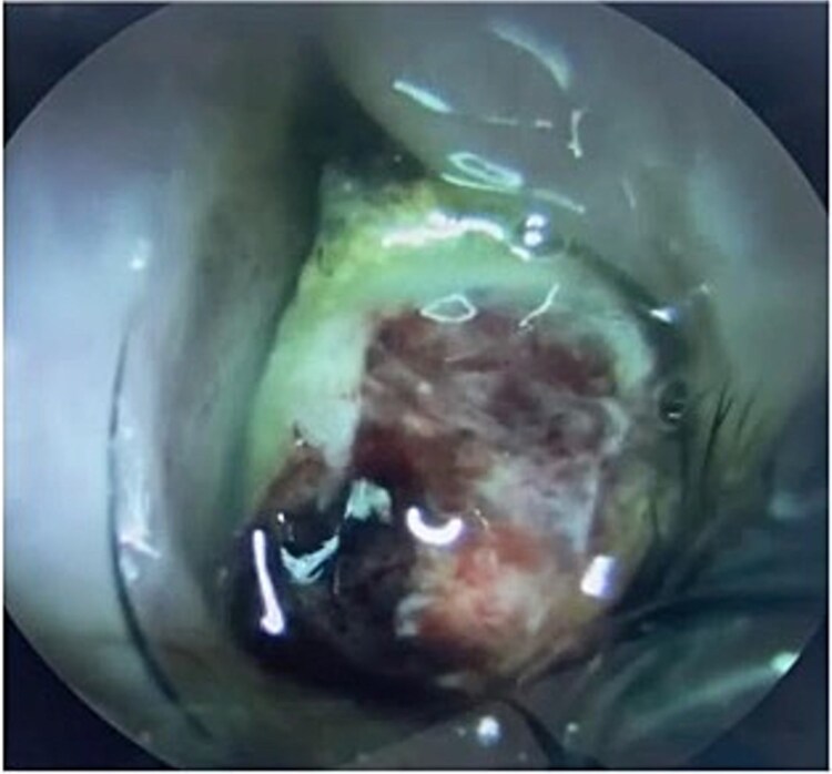
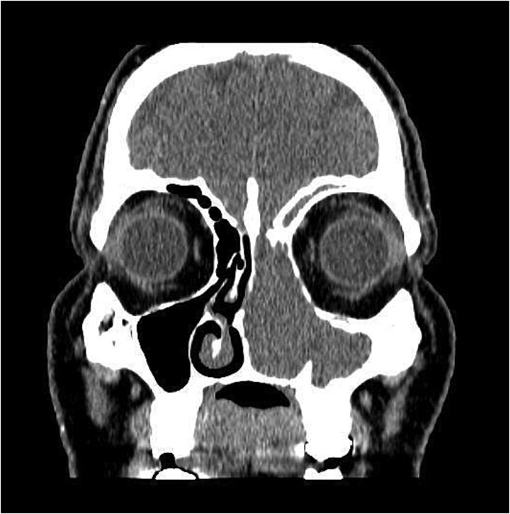
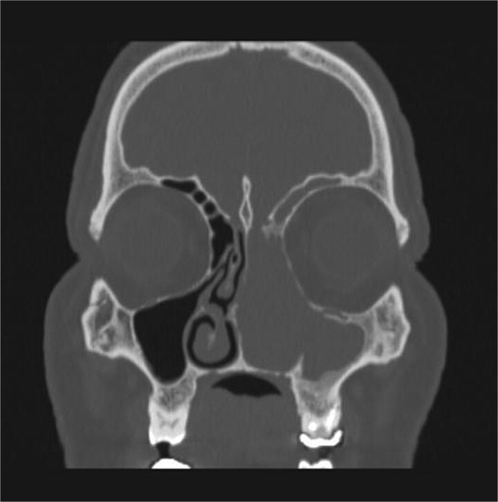
Sinonasal malignant melanoma (SNMM) is an extremely rare and aggressive sinonasal tumor. It has nonspecific risk factors and clinical presentation which require a high index of suspicion. In this case report we present a case of a 47-year-old male who initially had persistent nasal obstruction not responding to intranasal steroids. Comprehensive imaging and histopathological studies confirmed SNMM. He underwent incomplete maxillectomy resection followed by immunotherapy abroad. Within 6 months period he had recurrence of the disease. As this disease is a rare entity, there are no specific treatment guidelines, however, surgical intervention is agreed to be the mainstay of treatment. Although their effectiveness is still controversial, radiation therapy, chemotherapy, immunological and biological therapies are adjuvant options. Multidisciplinary team approach is necessary to provide the optimal treatment plan, along with close follow up. Continuous research and clinical trials are still needed to ensure appropriate diagnosis and management of this rare entity.
Keywords: malignant melanoma; oncology; rhinology; sinonasal tumor; treatment guidelines.
Published by Oxford University Press and JSCR Publishing Ltd. © The Author(s) 2025.
Conflict of interest statement
None declared.
Figures
Similar articles
-
Ocular invasion in sinonasal malignant melanoma: A case report and review of literature.Int J Surg Case Rep. 2025 Feb;127:110904. doi: 10.1016/j.ijscr.2025.110904. Epub 2025 Jan 20. Int J Surg Case Rep. 2025. PMID: 39842280 Free PMC article.
-
Vesicoureteral Reflux.2024 Apr 30. In: StatPearls [Internet]. Treasure Island (FL): StatPearls Publishing; 2025 Jan–. 2024 Apr 30. In: StatPearls [Internet]. Treasure Island (FL): StatPearls Publishing; 2025 Jan–. PMID: 33085409 Free Books & Documents.
-
Nasal Malignant Melanoma With an Inverted Papilloma in the Contralateral Nasal Cavity: A Case Report.Ear Nose Throat J. 2023 Jun 8:1455613231179692. doi: 10.1177/01455613231179692. Online ahead of print. Ear Nose Throat J. 2023. PMID: 37291873
-
Sinonasal mucosal melanoma: treatment strategies and survival rates for a rare disease entity : A single center experience and review of literature.Wien Klin Wochenschr. 2021 Nov;133(21-22):1137-1147. doi: 10.1007/s00508-021-01847-6. Epub 2021 Apr 12. Wien Klin Wochenschr. 2021. PMID: 33844113 Free PMC article. Review.
-
Contemporary Multidisciplinary Management of Sinonasal Mucosal Melanoma.Onco Targets Ther. 2020 Mar 16;13:2289-2298. doi: 10.2147/OTT.S182580. eCollection 2020. Onco Targets Ther. 2020. PMID: 32214828 Free PMC article. Review.
References
-
- Mihajlovic M, Vlajkovic S, Jovanovic P, et al. Primary mucosal melanomas: a comprehensive review. Int J Clin Exp Pathol 2012;5:739–53. Available from https://pmc.ncbi.nlm.nih.gov/articles/PMC3466987/. - PMC - PubMed
Publication types
LinkOut - more resources
Full Text Sources
Research Materials
