. 2025 Mar:110:1-110.
doi: 10.3114/sim.2025.110.01. Epub 2024 Dec 20.

An integrative re-evaluation of the Fusarium sambucinum species complex

Affiliations

An integrative re-evaluation of the Fusarium sambucinum species complex

M Sandoval-Denis et al. Stud Mycol. 2025 Mar.

Abstract

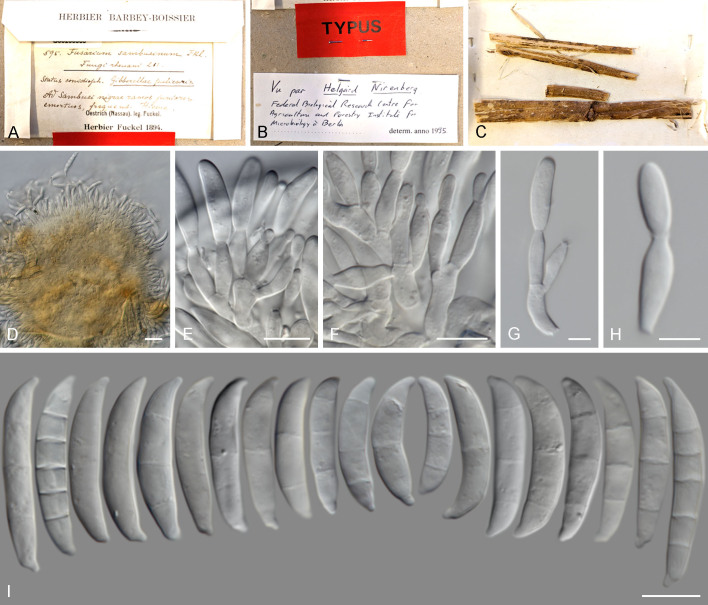
The species-rich Fusarium sambucinum species complex (FSAMSC; Fusarium, Nectriaceae, Hypocreales) is well-known for including devastating plant pathogens and toxigenic species. However, this group of grass-loving fungi also accommodates soil saprobes, endophytes, mycoparasites and rare opportunistic pathogens of humans and other animals. Recent publications have highlighted the vast phylogenetic and biochemical diversity of the FSAMSC, although a large number of taxa in FSAMSC have not been systematically described and still lack Latin binomials. In this study we established the phylogenetic breadth of the FSAMSC using an integrative approach including morphological, multilocus phylogenetic, and coalescence analyses based on five gene regions (calmodulin, RNA polymerase II largest and second largest subunits, translation elongation factor 1-α, and β-tubulin). Results obtained support the recognition of 75 taxa in FSAMSC, including all the currently known species segregates of the Fusarium head-blight pathogen F. graminearum s. lat. Thirty novel species are formally described and illustrated, while four phylogenetic species remain undescribed. An epitype is proposed for the generic type of Fusarium, F. sambucinum, from recently collected material identified by means of morphology, phylogenetics and mating experiments, fixing the phylogenetic application of the name. Additional notes are included on the typification of Fusisporium cerealis (syn. Fusarium cerealis). Taxonomic novelties: New species: Fusarium agreste Sand.-Den., J.Z. Groenew. & Crous, Fusarium amblysporum Sand.-Den., M.M. Costa, Fusarium bananae Sand.-Den., M.M. Costa, Fusarium bellum Sand.-Den., J.Z. Groenew. & Crous, Fusarium brachypes Sand.-Den., J.Z. Groenew. & Crous, Fusarium carinatum Sand.-Den., J.Z. Groenew. & Crous, Fusarium cultriforme Sand.-Den., M.M. Costa, Fusarium cuspidatum Sand.-Den., J.Z. Groenew. & Crous, Fusarium cygneum Sand.-Den., J.Z. Groenew. & Crous, Fusarium dimorphosporum Sand.-Den., M.M. Costa, J.Z. Groenew. & Crous, Fusarium dolichosporum Sand.-Den., J.Z. Groenew. & Crous, Fusarium gladiolum Sand.-Den., J.Z. Groenew. & Crous, Fusarium hamatum Sand.-Den., M.M. Costa, J.Z. Groenew. & Crous, Fusarium leptum Sand.-Den., J.Z. Groenew. & Crous, Fusarium longicolle Sand.-Den., J.Z. Groenew. & Crous, Fusarium magnum Sand.-Den., J.Z. Groenew. & Crous, Fusarium mastigosporum Sand.-Den., M.M. Costa, J.Z. Groenew. & Crous, Fusarium minutum Sand.-Den., J.Z. Groenew. & Crous, Fusarium mucronatum Sand.-Den., J.Z. Groenew. & Crous, Fusarium parabolicum Sand.-Den., J.Z. Groenew. & Crous, Fusarium platysporum Sand.-Den., J.Z. Groenew. & Crous, Fusarium pratense Sand.-Den., J.Z. Groenew. & Crous, Fusarium procumbens Sand.-Den., J.Z. Groenew. & Crous, Fusarium pseudolongipes Sand.-Den., J.Z. Groenew. & Crous, Fusarium sagittatum Sand.-Den., J.Z. Groenew. & Crous, Fusarium seculiforme Sand.-Den., J.Z. Groenew. & Crous, Fusarium subcylindroides Sand.-Den., J.Z. Groenew. & Crous, Fusarium symmetricum Sand.-Den., J.Z. Groenew. & Crous, Fusarium tropicale Sand.-Den., M.M. Costa, J.Z. Groenew. & Crous, Fusarium vermicularioides Sand.-Den., J.Z. Groenew. & Crous. Epitype: Fusarium sambucinum Fuckel. Citation: Sandoval-Denis M, Costa MM, Broders K, Becker Y, Maier W, Yurkov A, Kermode A, Buddie AG, Ryan MJ, Schumacher RK, Groenewald JZ, Crous PW (2024). An integrative re-evaluation of the Fusarium sambucinum species complex. Studies in Mycology 110: 1-110 doi: 10.3114/sim.2025.110.01.

Keywords: Coalescence; fungi; novel species; pathogens; phylogenetics; systematics; taxonomy.


Figures

Fig. 1
Fig. 1
Maximum likelihood (IQ-TREE) phylogeny of the Fusarium sambucinum species complex (FSAMSC) inferred from combined, partial CaM, rpb1, rpb2, tef1, and tub2 loci. The tree is rooted to Fusarium circinatum CBS 405.97 (F. fujikuroi species complex). Numbers at the nodes are IQ-TREE ultra-fast bootstrap (BS) (UFBoot) values ≥ 95 %, followed by RAxML BS ≥ 70 %, and MrBayes posterior probability (PP) ≥0. 95. Full supported branches (UFBoot and BS = 100 %, and PP = 1) are indicated in bold. Strain codes in bold correspond to type strains; and ex-epitype, ex-isotype, ex-neotype, and ex-type strains are indicated with ET, IT, NT, and T, respectively. Strains accessions are followed by substrate/host information (in red), and geographic origin (in blue). Names of novel taxa described in this paper are in bold.
Fig. 1
Fig. 1
Maximum likelihood (IQ-TREE) phylogeny of the Fusarium sambucinum species complex (FSAMSC) inferred from combined, partial CaM, rpb1, rpb2, tef1, and tub2 loci. The tree is rooted to Fusarium circinatum CBS 405.97 (F. fujikuroi species complex). Numbers at the nodes are IQ-TREE ultra-fast bootstrap (BS) (UFBoot) values ≥ 95 %, followed by RAxML BS ≥ 70 %, and MrBayes posterior probability (PP) ≥0. 95. Full supported branches (UFBoot and BS = 100 %, and PP = 1) are indicated in bold. Strain codes in bold correspond to type strains; and ex-epitype, ex-isotype, ex-neotype, and ex-type strains are indicated with ET, IT, NT, and T, respectively. Strains accessions are followed by substrate/host information (in red), and geographic origin (in blue). Names of novel taxa described in this paper are in bold.
Fig. 2
Fig. 2
Distributions of overall macroconidial sizes of species in FSAMSC. Average sizes are indicated by white dashed lines. Typical macroconidial morphology for each species is shown in the right-most column (not up to scale). Only mesoconidia produced. No sporodochial conidia produced.
Fig. 3
Fig. 3
Summary of species delimitation results by phylogenetic and coalescent methods of a reduced dataset to minimize missing data. Analyses referred to as “Combined” and STACEY (Species Tree And Classification Estimation) are based on multilocus datasets. Phylo = single-locus phylogeny, ABGD = Automatic Barcode Gap Discovery, ASAP = Assemble Species by Automatic Partitioning, bPTP = Bayesian implementation of the Poisson Tree Process model. Samples are split and colour coded according to hypothetical species and monophyletic clades and mapped against the result of a 5-gene IQ-TREE multilocus phylogenetic analysis. Blank spaces correspond to missing sequence data for the respective strain. Type strains are indicated in bold, followed by ET, IT, NT, and T to denote, respectively, ex-epitype, ex-isotype, ex-neotype, and ex-type strains.
Fig. 3
Fig. 3
Summary of species delimitation results by phylogenetic and coalescent methods of a reduced dataset to minimize missing data. Analyses referred to as “Combined” and STACEY (Species Tree And Classification Estimation) are based on multilocus datasets. Phylo = single-locus phylogeny, ABGD = Automatic Barcode Gap Discovery, ASAP = Assemble Species by Automatic Partitioning, bPTP = Bayesian implementation of the Poisson Tree Process model. Samples are split and colour coded according to hypothetical species and monophyletic clades and mapped against the result of a 5-gene IQ-TREE multilocus phylogenetic analysis. Blank spaces correspond to missing sequence data for the respective strain. Type strains are indicated in bold, followed by ET, IT, NT, and T to denote, respectively, ex-epitype, ex-isotype, ex-neotype, and ex-type strains.
Fig. 4
Fig. 4
Results of STACEY species delimitations of the Brachygibbosum clade of FSAMSC. Similarity matrix showing the probability (Bayesian posterior probabilities, PP) of each strain pair to belong to the same species group. Intensity of coloured blocks is equivalent to PP values, where white = 0 and dark green = 1. The matrix is mapped to a maximum credibility tree of the Brachygibbosum clade generated in BEAST, based on combined CaM, rpb1, rpb2, tef1, and tub2 loci. Values at the nodes indicate PP ≥ 0.95. Branches in bold indicate PP = 1. Ex-type strains are indicated by T and bold font. The inset shows the position of the Brachygibbosum clade with respect to the phylogeny of the entire FSAMSC as shown in Fig. 1
Fig. 5
Fig. 5
Results of STACEY species delimitations of the Graminearum clade of FSAMSC. Similarity matrix showing the probability (Bayesian posterior probabilities, PP) of each strain pair to belong to the same species group. Intensity of coloured blocks is equivalent to PP values, where white = 0 and dark green = 1. The matrix is mapped to a maximum credibility tree of the Graminearum clade generated in BEAST, based on combined CaM, rpb1, rpb2, tef1, and tub2 loci. Values at the nodes indicate PP ≥ 0.95. Branches in bold indicate PP = 1. Ex-epitype, and ex-type strains are indicated by ET, and T, respectively; and bold font. The inset shows the position of the Graminearum clade with respect to the phylogeny of the entire FSAMSC as shown in Fig. 1.
Fig. 6
Fig. 6
Results of STACEY species delimitations of the Longipes clade of FSAMSC. Similarity matrix showing the probability (Bayesian posterior probabilities, PP) of each strain pair to belong to the same species group. Intensity of coloured blocks is equivalent to PP values, where white = 0 and dark green = 1. The matrix is mapped to a maximum credibility tree of the Longipes clade generated in BEAST, based on combined CaM, rpb1, rpb2, tef1, and tub2 loci. Values at the nodes indicate PP ≥ 0.95. Branches in bold indicate PP = 1. Ex-neotype, and ex-type strains are indicated by NT, and T, respectively; and bold font. The inset shows the position of the Longipes clade with respect to the phylogeny of the entire FSAMSC as shown in Fig. 1.
Fig. 7
Fig. 7
Results of STACEY species delimitations of the Sambucinum clade of FSAMSC. Similarity matrix showing the probability (Bayesian posterior probabilities, PP) of each strain pair to belong to the same species group. Intensity of coloured blocks is equivalent to PP values, where white = 0 and dark green = 1. The matrix is mapped to a maximum credibility tree of the Sambucinum clade generated in BEAST, based on combined CaM, rpb1, rpb2, tef1, and tub2 loci. Values at the nodes indicate PP ≥ 0.95. Branches in bold indicate PP = 1. Ex-epitype, ex-isotype, and ex-type strains are indicated by ET, IT, and T, respectively; and bold font. The inset shows the position of the Sambucinum clade with respect to the phylogeny of the entire FSAMSC as shown in Fig. 1.
Fig. 8
Fig. 8
Results of STACEY species delimitations of the Gladiolum and Sporotrichioides clades of FSAMSC. Similarity matrix showing the probability (Bayesian posterior probabilities, PP) of each strain pair to belong to the same species group. Intensity of coloured blocks is equivalent to PP values, where white = 0 and dark green = 1. The matrix is mapped to a maximum credibility tree of the Gladiolum and Sporotrichioides clades generated in BEAST, based on combined CaM, rpb1, rpb2, tef1, and tub2 loci. Values at the nodes indicate PP ≥ 0.95. Branches in bold indicate PP = 1. Ex-epitype, and ex-type strains are indicated by ET, and T, respectively; and bold font. The inset shows the position of the Gladiolum and Sporotrichioides clades with respect to the phylogeny of the entire FSAMSC as shown in Fig. 1.
Fig. 9
Fig. 9
Fusarium acaciae-mearnsii (ex-type CBS 110254). A, B, D. Colonies on PDA, OA, and SNA respectively. C. Reverse of colonies on PDA (up), and OA (down). E–K. Conidiophores and conidiogenous cells. L. Macroconidia. Scale bars: E–I, L = 10 μm; J, K = 5 μm .
Fig. 10
Fig. 10
Fusarium aethiopicum (ex-type CBS 122858). A, B, D. Colonies on PDA, OA, and SNA respectively. C. Reverse of colonies on PDA (up), and OA (down). E. Sporodochia. F. Sporodochial conidiophores and conidiogenous cells. G, H. Aerial conidiophores and conidiogenous cells. I. Microconidia. J. Macroconidia. Scale bars = 10 μm.
Fig. 11
Fig. 11
Fusarium agreste (ex-type CBS 120993). A, B, D. Colonies on PDA, OA, and SNA respectively. C. Reverse of colonies on PDA (up), and OA (down). E, F. Sporodochia. G, H. Aerial conidiogenous cells. I. Microconidia. J. Sporodochial conidiophores and conidiogenous cells. K. Macroconidia. Scale bars: G, H = 5 μm; I–K = 10 μm.
Fig. 12
Fig. 12
Fusarium amblysporum (ex-type CBS 151809). A, B, D. Colonies on PDA, OA, and SNA respectively. C. Reverse of colonies on PDA (up), and OA (down). E. Sporodochia. F. Sporodochial conidiophore and conidiogenous cells. G, H. Chlamydospores. I. Aerial conidiogenous cells. J. Macroconidia. Scale bars = 10 μm.
Fig. 13
Fig. 13
Fusarium armeniacum (ex-type CBS 485.94). A, B, D. Colonies on PDA, OA, and SNA respectively. C. Reverse of colonies on PDA (up), and OA (down). E. Sporodochia. F. Sporodochial conidiophore and conidiogenous cells. G. Aerial conidiophore and conidiogenous cells. H. Microconidia. I. Macroconidia. Scale bars = 10 μm.
Fig. 14
Fig. 14
Fusarium asiaticum (ex-type CBS 110257). A, B, D. Colonies on PDA, OA, and SNA respectively. C. Reverse of colonies on PDA (up), and OA (down). E. Sporodochia. F, G. Sporodochial conidiophores and conidiogenous cells. H, I. Aerial conidiogenous cells. J. Macroconidia. Scale bars: F, H, J = 10 μm; G, I = 5 μm.
Fig. 15
Fig. 15
Fusarium austroamericanum (ex-type CBS 110244). A, B, D. Colonies on PDA, OA, and SNA respectively. C. Reverse of colonies on PDA (up), and OA (down). E–H. Sporodochial conidiophores and conidiogenous cells. I. Macroconidia. Scale bars = 10 μm.
Fig. 16
Fig. 16
Fusarium austroamericanum sexual morph (CBS 110245). A, B. Perithecia. C. Detail of perithecial wall cells. D, E. Asci. F. Ascospores. Scale bars: A, B = 100 μm;, C–F = 10 μm.
Fig. 17
Fig. 17
Fusarium bananae (ex-type CBS 151937). A, B, D. Colonies on PDA, OA, and SNA respectively. C. Reverse of colonies on PDA (up), and OA (down). E–G. Sporodochia. H–J. Sporodochial conidiophores and conidiogenous cells. K–M. Aerial conidiophores and conidiogenous cells. N. Macroconidia. Scale bars = 10 μm.
Fig. 18
Fig. 18
Fusarium bellum (ex-type CBS 151827). A, B, D. Colonies on PDA, OA, and SNA respectively. C. Reverse of colonies on PDA (up), and OA (down). E. Sporodochia. F. Sporodochial conidiophore and conidiogenous cells. G. Aerial conidiophores and conidiogenous cells. H. Microconidia. I. Chlamydospores. J. Macroconidia. Scale bars: F, G, I, J = 10 μm; H = 5 μm.
Fig. 19
Fig. 19
Fusarium boothii (ex-type CBS 316.73). A, B, D. Colonies on PDA, OA, and SNA respectively. C. Reverse of colonies on PDA (up), and OA (down). E. Sporodochia. F, G. Sporodochial conidiophores and conidiogenous cells. H–J. Aerial conidiogenous cells. K. Macroconidia. Scale bars = 10 μm.
Fig. 20
Fig. 20
Fusarium brachygibbosum (ex-type NRRL 20954). A, B, D. Colonies on PDA, OA, and SNA respectively. C. Reverse of colonies on PDA (up), and OA (down). E–I. Conidiogenous cells. J. Chlamydospore. K. Microconidia. L. Macroconidia. Scale bars: E–G, J–L = 10 μm; H, I = 5 μm.
Fig. 21
Fig. 21
Fusarium brachypes (ex-type CBS 151824). A, B, D. Colonies on PDA, OA, and SNA respectively. C. Reverse of colonies on PDA (up), and OA (down). E. Cluster of macroconidia on agar surface. F–I. Aerial conidiophores and conidiogenous cells. J. Microconidia. K. Macroconidia. Scale bars = 10 μm.
Fig. 22
Fig. 22
Fusarium carinatum (ex-type CBS 151957). A, B, D. Colonies on PDA, OA, and SNA respectively. C. Reverse of colonies on PDA (up), and OA (down). E, F. Sporodochia. G. Sporodochial conidiophores and conidiogenous cells. H, I. Aerial conidiophores, conidiogenous cells and microconidia. J. Aerial macroconidia. K. Chlamydospores. L. Sporodochial macroconidia. Scale bars = 10 μm.
Fig. 23
Fig. 23
Fusarium cerealis (neotype K(M)133541). A. Specimen label. B. Specimen. C, D. Macroconidia from specimen as depicted in Nirenberg (1990). E. Microphotograph of macroconidia. F. Microphotograph of sporodochial conidiophore and conidiogenous cells. G. Macroconidia. Scale bar = 10 μm.
Fig. 24
Fig. 24
Fusarium cerealis (CBS 832.85). A, B, D. Colonies on PDA, OA, and SNA respectively. C. Reverse of colonies on PDA (up), and OA (down). E, F. Sporodochia. G, H. Sporodochial conidiophores and conidiogenous cells. I. Aerial conidiophores. J. Macroconidia. Scale bars = 10 μm.
Fig. 25
Fig. 25
Fusarium chaquense (NRRL 64072). A, B, D. Colonies on PDA, OA, and SNA respectively. C. Reverse of colonies on PDA (up), and OA (down). E, F. Sporodochia. G, H. Sporodochial conidiophores and conidiogenous cells. I. Aerial conidiophores, conidiogenous cells and microconidia. J. Macroconidia. Scale bars = 10 μm.
Fig. 26
Fig. 26
Fusarium cortaderiae (ex-epitype CBS 119183). A, B, D. Colonies on PDA, OA, and SNA respectively. C. Reverse of colonies on PDA (up), and OA (down). E. Sporodochia. F. Sporodochial conidiogenous cells. G, H. Aerial conidiophores and conidiogenous cells. I. Macroconidia. Scale bars = 10 μm.
Fig. 27
Fig. 27
Fusarium culmorum (lectotype of Fusisporium culmorum). Reproduction from the original publication by Smith (1884). A–C. Conidiogenous cells and macroconidia. D. Host cells (wheat).
Fig. 28
Fig. 28
Fusarium culmorum (ex-epitype CBS 417.86). A, B, D. Colonies on PDA, OA, and SNA respectively. C. Reverse of colonies on PDA (up), and OA (down). E, F. Sporodochia. G, H. Sporodochial conidiogenous cells. I, J. Aerial conidiogenous cells. K. Macroconidia undergoing microcyclic conidiogenesis. L. Macroconidia. Scale bars: G–J = 5 μm; K, L = 10 μm.
Fig. 29
Fig. 29
Fusarium cultriforme (ex-type CBS 151935). A, B, D. Colonies on PDA, OA, and SNA respectively. C. Reverse of colonies on PDA (up), and OA (down). E, F. Sporodochia. G, H. Aerial conidiophores and conidiogenous cells. I. Sporodochial conidiophore and conidiogenous cells. J. Microconidia. K. Macroconidia. Scale bars: G–I, K = 10 μm; J = 5 μm.
Fig. 30
Fig. 30
Fusarium cuspidatum (ex-type CBS 151953). A, B, D. Colonies on PDA, OA, and SNA respectively. C. Reverse of colonies on PDA (up), and OA (down). E, F. Sporodochia. G, H. Sporodochial conidiophores and conidiogenous cells. I. Aerial conidiogenous cells. J. Macroconidia. Scale bars = 10 μm.
Fig. 31
Fig. 31
Fusarium cygneum (ex-type CBS 151805). A, B, D. Colonies on PDA, OA, and SNA respectively. C. Reverse of colonies on PDA (up), and OA (down). E, F. Sporodochia. G, H. Sporodochial conidiophores and conidiogenous cells. I. Aerial conidiogenous cells. J. Macroconidia. Scale bars = 10 μm.
Fig. 32
Fig. 32
Fusarium dactylidis (ex-type CBS 119181). A, B, D. Colonies on PDA, OA, and SNA respectively. C. Reverse of colonies on PDA (up), and OA (down). E–L. Conidiophores and conidiogenous cells. M. Macroconidia. Scale bars = 10 μm.
Fig. 33
Fig. 33
Fusarium dimorphosporum (ex-type CBS 151959). A, B, D. Colonies on PDA, OA, and SNA respectively. C. Reverse of colonies on PDA (up), and OA (down). E, G, H, J. Conidiophores and conidiogenous cells. F. Chlamydospores. I. Microconidia. K. Aerial macroconidia. L. Sporodochial macroconidia. Scale bars = 10 μm.
Fig. 34
Fig. 34
Fusarium dolichosporum (ex-type CBS 151817). A, B, D. Colonies on PDA, OA, and SNA respectively. C. Reverse of colonies on PDA (up), and OA (down). E–H. Conidiophores and conidiogenous cells. I. Microconidia. J. Macroconidia. Scale bars = 10 μm.
Fig. 35
Fig. 35
Fusarium gerlachii (ex-type CBS 119175). A, B, D. Colonies on PDA, OA, and SNA respectively. C. Reverse of colonies on PDA (up), and OA (down). E–G. Sporodochia. H–J. Sporodochial conidiophores and conidiogenous cells. K. Aerial conidiophore and conidiogenous cell. L. Macroconidia. Scale bars = 10 μm.
Fig. 36
Fig. 36
Fusarium gladiolum (ex-type CBS 151812). A, B, D. Colonies on PDA, OA, and SNA respectively. C. Reverse of colonies on PDA (up), and OA (down). E. Sporodochia. F, G. Sporodochial conidiophores and conidiogenous cells. H, I. Aerial conidiophores and conidiogenous cells. J. Macroconidia. Scale bars = 10 μm.
Fig. 37
Fig. 37
Fusarium goolgardi (ex-type CBS 151815). A, B, D. Colonies on PDA, OA, and SNA respectively. C. Reverse of colonies on PDA (up), and OA (down). E. Sporodochia. F. Sporodochial conidiophore and conidiogenous cells. G, H. Aerial conidiophores and conidiogenous cells. I. Microconidia. J. Macroconidia. Scale bars = 10 μm.
Fig. 38
Fig. 38
Fusarium graminearum (lectotype). Reproduction from the original publication by Schwabe (1884). A, B. Macroconidia.
Fig. 39
Fig. 39
Fusarium graminearum (ex-epitype CBS 136009). A, B, D. Colonies on PDA, OA, and SNA respectively. C. Reverse of colonies on PDA (up), and OA (down). E, F. Sporodochia. G, H. Sporodochial conidiophores and conidiogenous cells. I. Aerial conidiogenous cell. J. Macroconidia. Scale bars = 10 μm.
Fig. 40
Fig. 40
Fusarum hamatum (ex-type CBS 151936). A, B, D. Colonies on PDA, OA, and SNA respectively. C. Reverse of colonies on PDA (up), and OA (down). E. Sporodochia. F, H. Aerial conidiophores and conidiogenous cells. G. Chlamydospores. I. Sporodochial conidiophore and conidiogenous cells. J. Macroconidia. Scale bars = 10 μm.
Fig. 41
Fig. 41
Fusarium kyushuense (ex-type CBS 151828). A, B, D. Colonies on PDA, OA, and SNA respectively. C. Reverse of colonies on PDA (up), and OA (down). E–I. Conidiophores and conidiogenous cells. J. Microconidia. K. Macroconidia. Scale bars = 10 μm.
Fig. 42
Fig. 42
Fusarium langsethiae (ex-type CBS 113234). A, B, D. Colonies on PDA, OA, and SNA respectively. C. Reverse of colonies on PDA (up), and OA (down). E–H. Conidiophores and conidiogenous cells. I. Microconidia. Scale bars = 10 μm.
Fig. 43
Fig. 43
Fusarium leptum (ex-type CBS 151949). A, B, D. Colonies on PDA, OA, and SNA respectively. C. Reverse of colonies on PDA (up), and OA (down). E. Sporodochium. F, G. Sporodochial conidiophores and conidiogenous cells. H–J. Aerial conidiophores and conidiogenous cells. K. Macroconidia. Scale bars: F, G, K = 10 μm; I, J = 5 μm.
Fig. 44
Fig. 44
Fusarium longicolle (ex-type CBS 151818). A, B, D. Colonies on PDA, OA, and SNA respectively. C. Reverse of colonies on PDA (up), and OA (down). E, F. Sporodochia. G. Sporodochial conidiophores and conidiogenous cells. H, I. Aerial conidiophores and conidiogenous cells. J. Macroconidia. Scale bars: G, H, J = 10 μm; I = 5 μm.
Fig. 45
Fig. 45
Fusarium longipes (ex-neotype CBS 476.77). A, B, D. Colonies on PDA, OA, and SNA respectively. C. Reverse of colonies on PDA (up), and OA (down). E, F. Sporodochia. G. Sporodochial conidiophores and conidiogenous cells. H–J. Aerial conidiogenous cells. K. Aerial macroconidia. L. Sporodochial macroconidia. Scale bars: G, K, L = 10 μm; H–J = 5 μm.
Fig. 46
Fig. 46
Fusarium louisianense (ex-type CBS 127525). A, B, D. Colonies on PDA, OA, and SNA respectively. C. Reverse of colonies on PDA (up), and OA (down). E. Sporodochia. F. Sporodochial conidiophore and conidiogenous cells. G, H. Aerial conidiophores and conidiogenous cells. I. Macroconidia. Scale bars = 10 μm.
Fig. 47
Fig. 47
Fusarium lunulosporum (ex-type CBS 636.76). A, B, D. Colonies on PDA, OA, and SNA respectively. C. Reverse of colonies on PDA (up), and OA (down). E. Sporodochium. F, G. Sporodochial conidiophores and conidiogenous cells. H. Aerial conidiogenous cells. I. Macroconidia. Scale bars = 10 μm.
Fig. 48
Fig. 48
Fusarium magnum (ex-type CBS 151821). A, B, D. Colonies on PDA, OA, and SNA respectively. C. Reverse of colonies on PDA (up), and OA (down). E–G. Sporodochia. H. Sporodochial conidiophore and conidiogenous cells. I. Aerial conidiogenous cells. J. Macroconidia. Scale bars = 10 μm.
Fig. 49
Fig. 49
Fusarium mastigosporum (ex-type CBS 151825). A, B, D. Colonies on PDA, OA, and SNA respectively. C. Reverse of colonies on PDA (up), and OA (down). E. Sporodochium. F. Sporodochial conidiophores and conidiogenous cells. G–I. Aerial conidiophores and conidiogenous cells. J. Aerial macroconidia. K. Sporodochial macroconidia. Scale bars: F, H, J, K = 10 μm; I = 5 μm.
Fig. 50
Fig. 50
Fusarium meridionale (ex-type CBS 110247). A, B, D. Colonies on PDA, OA, and SNA respectively. C. Reverse of colonies on PDA (up), and OA (down). E. Sporodochia. F. Sporodochial conidiophore and conidiogenous cells. G–I. Aerial conidiophores and conidiogenous cells. J. Macroconidia. Scale bars = 10 μm.
Fig. 51
Fig. 51
Fusarium mesoamericanum (ex-type CBS 415.86). A, B, D. Colonies on PDA, OA, and SNA respectively. C. Reverse of colonies on PDA (up), and OA (down). E, F. Sporodochia. G. Sporodochial conidiophore and conidiogenous cells. H. Aerial conidiophore. I. Macroconidia. Scale bars = 10 μm.
Fig. 52
Fig. 52
Fusarium minutum (ex-type CBS 151823). A, B, D. Colonies on PDA, OA, and SNA respectively. C. Reverse of colonies on PDA (up), and OA (down). E–I. Conidiophores and conidiogenous cells. J. Microconidia. K. Chlamydospores. L. Macroconidia. Scale bars: E, H–L = 10 μm; F, G = 5 μm.
Fig. 53
Fig. 53
Fusarium mucronatum (ex-type CBS 151826). A, B, D. Colonies on PDA, OA, and SNA respectively. C. Reverse of colonies on PDA (up), and OA (down). E, F. Sporodochia. G, H. Sporodochial conidiophores and conidiogenous cells. I. Aerial conidiophore and conidiogenous cells. J. Macroconidia. Scale bars = 10 μm.
Fig. 54
Fig. 54
Fusarium musarum (ex-type CBS 151808). A, B, D. Colonies on PDA, OA, and SNA respectively. C. Reverse of colonies on PDA (up), and OA (down). E–H. Conidiophores and conidiogenous cells. I. Mesoconidium undergoing microcyclic conidiogenesis. J. Microconidia. K. Mesoconidia. Scale bars: E, F = 20 μm; G–K = 10 μm.
Fig. 55
Fig. 55
Fusarium palustre (ex-type CBS 126796). A, B, D. Colonies on PDA, OA, and SNA respectively. C. Reverse of colonies on PDA (up), and OA (down). E. Sporodochium and chlamydospores. F, G. Chlamydospores. H. Conidiogenous cell and microconidia. I–K. Conidiogenous cells. L. Macroconidia. Scale bars = 10 μm.
Fig. 56
Fig. 56
Fusarium parabolicum (ex-type CBS 151814). A, B, D. Colonies on PDA, OA, and SNA respectively. C. Reverse of colonies on PDA (up), and OA (down). E–G. Sporodochia. H. Sporodochial conidiophores and conidiogenous cells. I. Aerial conidiophore and conidiogenous cells. J. Chlamydospores. K. Microconidia. L. Macroconidia. Scale bars = 10 μm.
Fig. 57
Fig. 57
Fusarium platysporum (ex-type CBS 151816). A, B, D. Colonies on PDA, OA, and SNA respectively. C. Reverse of colonies on PDA (up), and OA (down). E, F. Sporodochia. G. Sporodochial conidiophores and conidiogenous cells. H. Aerial conidiophore and conidiogenous cell. I. Microconidia. J. Chlamydospores. K. Macroconidia. Scale bars: G, H, J, K = 10 μm; I = 5 μm.
Fig. 58
Fig. 58
Fusarium praegraminearum (ex-type CBS 141369). A, B, D. Colonies on PDA, OA, and SNA respectively. C. Reverse of colonies on PDA (up), and OA (down). E. Sporodochia. F. Sporodochial conidiophore and conidiogenous cells. G–I. Aerial conidiophores and conidiogenous cells. J. Macroconidia. Scale bars = 10 μm.
Fig. 59
Fig. 59
Fusarium pratense (ex-type CBS 151956). A, B, D. Colonies on PDA, OA, and SNA respectively. C. Reverse of colonies on PDA (up), and OA (down). E, F. Sporodochia. G–J. Aerial conidiophores and conidiogenous cells. K. Microconidia. L. Macroconidia. Scale bars = 10 μm.
Fig. 60
Fig. 60
Fusarium procumbens (ex-type CBS 121863). A, B, D. Colonies on PDA, OA, and SNA respectively. C. Reverse of colonies on PDA (up), and OA (down). E, F. Sporodochia. G, H. Aerial conidiophores and conidiogenous cells. I. Sporodochial conidiophore and conidiogenous cells. J. Macroconidia. Scale bars = 10 μm.
Fig. 61
Fig. 61
Fusarium pseudolongipes (ex-type CBS 131380). A, B, D. Colonies on PDA, OA, and SNA respectively. C. Reverse of colonies on PDA (up), and OA (down). E, F. Sporodochia. G, H. Sporodochial conidiophores and conidiogenous cells. I. Microconidia. J. Macroconidia. Scale bars = 10 μm.
Fig. 62
Fig. 62
Fusarium robustum (ex-isotype CBS 637.76). A, B, D. Colonies on PDA, OA, and SNA respectively. C. Reverse of colonies on PDA (up), and OA (down). E, F. Sporodochia. G, H. Sporodochial conidiophores and conidiogenous cells. I. Aerial conidiophore. J. Macroconidia. Scale bars = 10 μm.
Fig. 63
Fig. 63
Fusarium sagittatum (ex-type CBS 151958). A, B, D. Colonies on PDA, OA, and SNA respectively. C. Reverse of colonies on PDA (up), and OA (down). E–G. Conidiophores and conidiogenous cells. H. Chlamydospores. I. Microconidia. J. Macroconidia. Scale bars = 10 μm.
Fig. 64
Fig. 64
Fusarium sambucinum (lectotype G00266369). A–C. Herbarium specimen. D. Sporodochium. E–H. Sporodochial conidiophores and conidiogenous cells. I. Macroconidia. Scale bars: D = 20 μm; E, F, I = 10 μm; G, H = 5 μm.
Fig. 65
Fig. 65
Fusarium sambucinum (ex-epitype CBS 151942). A, B. Perithecia on host. C, D. Perithecia in culture. E–G. Detail of perithecial wall cells (G in 100 % lactic acid). H. Detail of perithecial anatomy (in 100 % lactic acid). I. Detail of ostiolar region (in 100 % lactic acid). J–L. Asci. M. Ascospores. N–P. Sporodochia on host. Q. Sporodochial conidiophores and conidiogenous cells. R. Micro- and macroconidia. S. Macroconidia. Scale bars: E (applies to F and G); H–K, Q = 20 μm; L, M, R, S = 10 μm.
Fig. 66
Fig. 66
Fusarium seculiforme (ex-type CBS 151806). A, B, D. Colonies on PDA, OA, and SNA respectively. C. Reverse of colonies on PDA (up), and OA (down). E. Sporodochia. F, G. Sporodochial conidiophores and conidiogenous cells. H. Aerial conidiophores and conidiogenous cells. I. Macroconidia. Scale bars = 10 μm.
Fig. 67
Fig. 67
Fusarium sibiricum (ex-type CBS 151951). A, B, D. Colonies on PDA, OA, and SNA respectively. C. Reverse of colonies on PDA (up), and OA (down). E–H. Conidiophores and conidiogenous cells. I. Microconidia. Scale bars = 10 μm.
Fig. 68
Fig. 68
Fusarium sporotrichioides (lectotype). Reproduction from the original publication by Sherbakoff (1915). A, E. Macroconidia. B, J, M, N. Aerial conidiophores. C, G. Balls of conidia. D. Germinating conidium. F. Chlamydospores. H. Sporodochial conidiophore. I, K, L. Microconidia. O. Plectenchymic stroma.
Fig. 69
Fig. 69
Fusarium sporotrichioides (CBS 448.67). A, B, D. Colonies on PDA, OA, and SNA respectively. C. Reverse of colonies on PDA (up), and OA (down). E. Sporodochium. F. Sporodochial conidiophore and conidiogenous cells. G–J. Aerial conidiophores and conidiogenous cells. K. Microconidia. L. Macroconidia. Scale bars = 10 μm.
Fig. 70
Fig. 70
Fusarium subcylindroides (ex-type CBS 151807). A, B, D. Colonies on PDA, OA, and SNA respectively. C. Reverse of colonies on PDA (up), and OA (down). E, F. Sporodochia. G–I. Sporodochial conidiophores and conidiogenous cells. J. Aerial conidiogenous cell. K. Macroconidia. Scale bars = 10 μm.
Fig. 71
Fig. 71
Fusarium subtropicale (ex-type CBS 144706). A, B, D. Colonies on PDA, OA, and SNA respectively. C. Reverse of colonies on PDA (up), and OA (down). E. Sporodochium. F. Sporodochial conidiophore and conidiogenous cells. G–J. Aerial conidiophores and conidiogenous cells. K. Macroconidia. Scale bars: F = 5 μm; G–K = 10 μm.
Fig. 72
Fig. 72
Fusarium symmetricum (ex-type CBS 151804). A, B, D. Colonies on PDA, OA, and SNA respectively. C. Reverse of colonies on PDA (up), and OA (down). E. Sporodochium. F, G. Sporodochial conidiophores and conidiogenous cells. H–J. Aerial conidiophores and conidiogenous cells. K. Macroconidia. Scale bars: F, G, I, K = 10 μm; J = 5 μm.
Fig. 73
Fig. 73
Fusarium tropicale (ex-type CBS 151820). A, B, D. Colonies on PDA, OA, and SNA respectively. C. Reverse of colonies on PDA (up), and OA (down). E, F. Sporodochia. G, H. Sporodochial conidiophores and conidiogenous cells. I, J. Aerial conidiophores and conidiogenous cells. K. Chlamydospores. L. Macroconidia. Scale bars: G, H, L = 10 μm; I–K = 5 μm.
Fig. 74
Fig. 74
Fusarium ussurianum (ex-type CBS 123752). A, B, D. Colonies on PDA, OA, and SNA respectively. C. Reverse of colonies on PDA (up), and OA (down). E, F. Sporodochia. G. Sporodochial conidiophore and conidiogenous cells. H, I. Aerial conidiogenous cells. J. Macroconidia. Scale bars = 10 μm.
Fig. 75
Fig. 75
Fusarium venenatum (ex-type CBS 458.93). A, B, D. Colonies on PDA, OA, and SNA respectively. C. Reverse of colonies on PDA (up), and OA (down). E. Sporodochia. F. Sporodochial conidiophore and conidiogenous cells. G–I. Aerial conidiophores and conidiogenous cells. J. Chlamydospores. K. Macroconidia. Scale bars: F–H, J, K = 10 μm; I = 5 μm.
Fig. 76
Fig. 76
Fusarium vermicularioides (ex-type CBS 151945). A, B, D. Colonies on PDA, OA, and SNA respectively. C. Reverse of colonies on PDA (up), and OA (down). E, F. Sporodochia. G. Sporodochial conidiophores and conidiogenous cells. H. Aerial conidiogenous cell. I. Aerial macroconidia and microconidium (arrow). J. Chlamydospores. K. Sporodochial macroconidia. Scale bars: G, I–K = 10 μm; H = 5 μm.
Fig. 77
Fig. 77
Fusarium vorosii (ex-type CBS 119177). A, B, D. Colonies on PDA, OA, and SNA respectively. C. Reverse of colonies on PDA (up), and OA (down). E. Sporodochia. F. Sporodochial conidiophore and conidiogenous cells. G, H. Aerial conidiogenous cells. I. Macroconidia. Scale bars = 10 μm.

Similar articles

  • Known from trees and the tropics: new insights into the Fusarium lateritium species complex.
    Costa MM, Sandoval-Denis M, Moreira GM, Kandemir H, Kermode A, Buddie AG, Ryan MJ, Becker Y, Yurkov A, Maier W, Groenewald JZ, Pfenning LH, Crous PW. Costa MM, et al. Stud Mycol. 2024 Dec;109:403-450. doi: 10.3114/sim.2024.109.06. Epub 2024 Sep 26. Stud Mycol. 2024. PMID: 39717659 Free PMC article.
  • Fusarium: more than a node or a foot-shaped basal cell.
    Crous PW, Lombard L, Sandoval-Denis M, Seifert KA, Schroers HJ, Chaverri P, Gené J, Guarro J, Hirooka Y, Bensch K, Kema GHJ, Lamprecht SC, Cai L, Rossman AY, Stadler M, Summerbell RC, Taylor JW, Ploch S, Visagie CM, Yilmaz N, Frisvad JC, Abdel-Azeem AM, Abdollahzadeh J, Abdolrasouli A, Akulov A, Alberts JF, Araújo JPM, Ariyawansa HA, Bakhshi M, Bendiksby M, Ben Hadj Amor A, Bezerra JDP, Boekhout T, Câmara MPS, Carbia M, Cardinali G, Castañeda-Ruiz RF, Celis A, Chaturvedi V, Collemare J, Croll D, Damm U, Decock CA, de Vries RP, Ezekiel CN, Fan XL, Fernández NB, Gaya E, González CD, Gramaje D, Groenewald JZ, Grube M, Guevara-Suarez M, Gupta VK, Guarnaccia V, Haddaji A, Hagen F, Haelewaters D, Hansen K, Hashimoto A, Hernández-Restrepo M, Houbraken J, Hubka V, Hyde KD, Iturriaga T, Jeewon R, Johnston PR, Jurjević Ž, Karalti I, Korsten L, Kuramae EE, Kušan I, Labuda R, Lawrence DP, Lee HB, Lechat C, Li HY, Litovka YA, Maharachchikumbura SSN, Marin-Felix Y, Matio Kemkuignou B, Matočec N, McTaggart AR, Mlčoch P, Mugnai L, Nakashima C, Nilsson RH, Noumeur SR, Pavlov IN, Peralta MP, Phillips AJL, Pitt JI, Polizzi G, Quaedvlieg W, Rajeshkumar KC, Restrepo S, Rhaiem A, Robert J, Robert V, Ro… See abstract for full author list ➔ Crous PW, et al. Stud Mycol. 2021 Aug 17;98:100116. doi: 10.1016/j.simyco.2021.100116. eCollection 2021 Apr. Stud Mycol. 2021. PMID: 34466168 Free PMC article.
  • Redisposition of acremonium-like fungi in Hypocreales.
    Hou LW, Giraldo A, Groenewald JZ, Rämä T, Summerbell RC, Huang GZ, Cai L, Crous PW. Hou LW, et al. Stud Mycol. 2023 Jun;105:23-203. doi: 10.3114/sim.2023.105.02. Epub 2023 Jun 2. Stud Mycol. 2023. PMID: 38895703 Free PMC article.
  • Towards an integrated phylogenetic classification of the Tremellomycetes.
    Liu XZ, Wang QM, Göker M, Groenewald M, Kachalkin AV, Lumbsch HT, Millanes AM, Wedin M, Yurkov AM, Boekhout T, Bai FY. Liu XZ, et al. Stud Mycol. 2015 Jun;81:85-147. doi: 10.1016/j.simyco.2015.12.001. Epub 2016 Jan 8. Stud Mycol. 2015. PMID: 26955199 Free PMC article.
  • Revising Clonostachys and allied genera in Bionectriaceae.
    Zhao L, Groenewald JZ, Hernández-Restrepo M, Schroers HJ, Crous PW. Zhao L, et al. Stud Mycol. 2023 Jun;105:205-266. doi: 10.3114/sim.2023.105.03. Epub 2023 Jun 12. Stud Mycol. 2023. PMID: 38895704 Free PMC article.

Cited by

References

    1. Akinsanmi OA, Chakraborty S, Backhouse D, et al. (2007). Passage through alternative hosts changes the fitness of Fusarium graminearum and Fusarium pseudograminearum. Environmental Microbiology 9: 512–520. - PubMed
    1. Al-Hatmi A. (2016). Phylogeny, diagnostics and antifungal susceptibility of clinically relevant Fusarium species. Ph.D. dissertation. Faculty of Science, Mathematics and Computer Science, University of Amsterdam, The Netherlands.
    1. Aoki T, O’Donnell K. (1998). Fusarium kyushuense sp. nov. from Japan. Mycoscience 39: 1–6.
    1. Aoki T, O’Donnell K. (1999a). Morphological and molecular characterization of Fusarium pseudograminearum sp. nov., formerly recognized as the Group 1 population of F. graminearum. Mycologia 91: 597–609.
    1. Aoki T, O’Donnell K. (1999b). Morphological characterization of Gibberella coronicola sp. nov., obtained through mating experiments of Fusarium pseudograminearum. Mycoscience 40: 443–453.

LinkOut - more resources