Review
. 2025 Apr 21;15(12):5666-5692.
doi: 10.7150/thno.109186. eCollection 2025.

Fish-derived biomaterials for tissue engineering: advances in scaffold fabrication and applications in regenerative medicine and cancer therapy

Affiliations
Review

Fish-derived biomaterials for tissue engineering: advances in scaffold fabrication and applications in regenerative medicine and cancer therapy

Seoyul Jo et al. Theranostics. .

Abstract

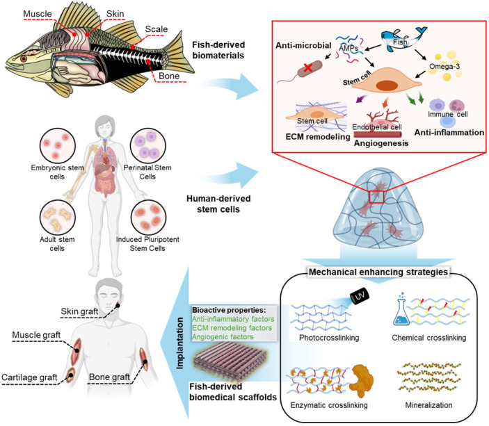
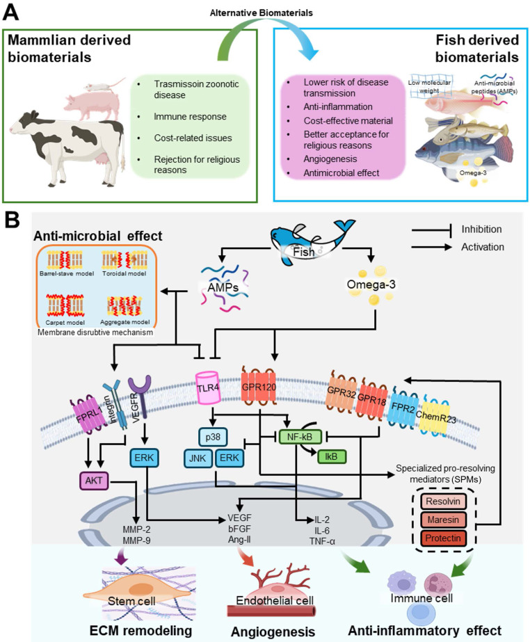
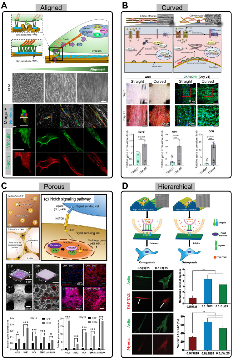
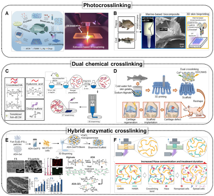
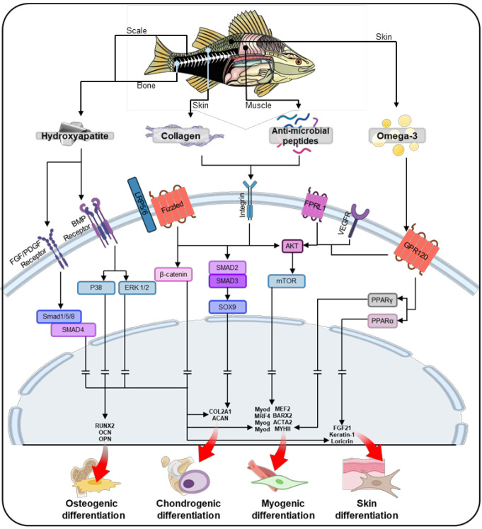
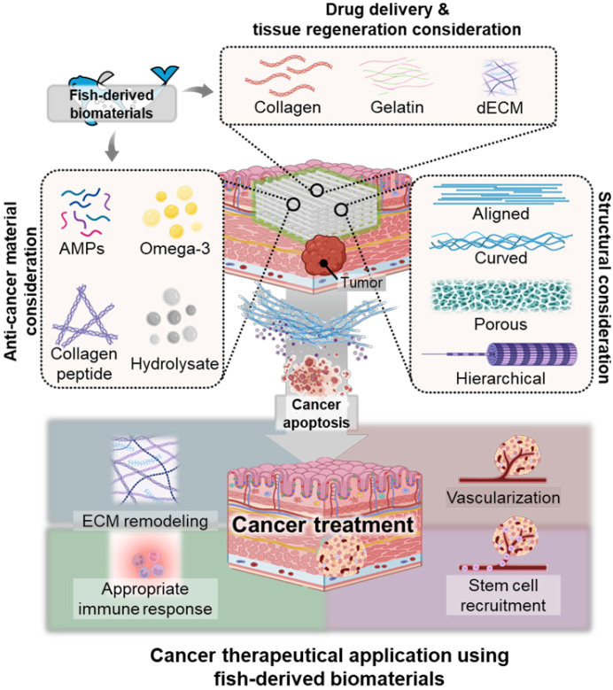
Fish-derived biomaterials, such as collagen, polyunsaturated fatty acids, and antimicrobial peptides, have emerged as promising candidates for scaffold development in stem cell therapies and tissue engineering due to their excellent biocompatibility and low immunogenicity. Although good bioactivity is a prerequisite for biomedical substitutes, scaffold design is necessary for the successful development of bioconstructs used in tissue regeneration. However, the limited processability of fish biomaterials poses a substantial challenge to the development of diverse scaffold structures. In this review, unlike previous reviews that primarily focused on the bioactivities of fish-derived components, we placed greater emphasis on scaffold fabrication and its applications in tissue regeneration. Specifically, we examined various cross-linking strategies to enhance the structural integrity of fish biomaterials and address challenges, such as poor processability, low mechanical strength, and rapid degradation. Furthermore, we demonstrated the potential of fish scaffolds in stem cell therapies, particularly their capacity to support stem cell growth and modulate the cellular microenvironment. Finally, this review provides future directions for the application of these scaffolds in cancer therapy.

Keywords: cancer therapeutics; fish-derived biomaterials; scaffold structure; stem cell activation; tissue engineering.


Conflict of interest statement

Competing Interests: The authors have declared that no competing interest exists.

Figures

Figure 1
Figure 1
Fish-derived biomaterials for scaffold fabrication in stem cell therapies: enhancing biocompatibility, supporting cell growth, and promoting tissue regeneration. ECM, extracellular matrix.
Figure 2
Figure 2
Bioactivities of stem cells cultured on fish-derived biomaterials. (A) Comparative analysis of mammalian and fish-derived biomaterials, and (B) intercellular pathways activated by fish-derived biomaterials that stimulate cellular responses, such as antimicrobial activity, extracellular matrix (ECM) remodeling, angiogenesis, and anti-inflammatory effects.
Figure 3
Figure 3
Stem cell interactions with various scaffold structures. (A) Schematic illustration showing how aligned substrates regulate the self-renewal process of human mesenchymal stem cells (hMSCs) (Adapted with permission from , copyright 2020). SEM images and immunofluorescence stainings of Vinculin and F-actin for various nanopatterned scaffolds. (B) Schematic of mechanotransduction pathways in straight and curved fibers (Adapted with permission from , copyright 2024). Images of Alizarin Red S (ARS) staining and DAPI/OPN, and relative expression of BMP2, OPN, and OCN in hASCs cultured on straight and curved fibers. (C) Illustration of the notch signaling pathway in highly porous scaffolds by stretching and active cell-to-cell interactions (Adapted with permission from , copyright 2022). Optical, SEM, DAPI/F-actin, and DAPI/OPN staining images of conventional scaffolds and collagen foam scaffolds, along with the relative gene expression of osteogenic genes and MAPK signaling genes (COL1, BMP2, OCN, ERK1/2, and p38 MAPK) at days 14 and 28. (D) Schematic showing of the interaction between hMSCs and different types of hierarchical structure (Adapted with permission from , copyright 2020). Immunofluorescence staining images of Actin, YAP-TAZ, and Myosin, along with fluorescence intensity of myosin and the number of cells showing nuclear localization of YAP-TAZ.
Figure 4
Figure 4
Crosslinking strategies to enhance mechanical capabilities of fish-derived biomedical scaffolds. (A) Schematics and optical image of the fabrication of the bio-printed fish-skin- decellularized extracellular matrix (dECM) methacrylate and application of skin regeneration (Adapted with permission from , copyright 2023). (B) Schematics of the bioprinting process of photocrosslinkable bioink composed of methacrylated tilapia dECM (tdECM-MA) and cod dECM (Adapted from , with permission). (C) Schematic of fish-derived dECM formulation and 3D hybrid bioink preparation (Adapted with permission from , 2023). Solubilized fish-dECM is pre-gelled using divinyl sulfone (DVS), followed by 3D printing to fabricate a structured model, with a secondary crosslinking step using DVS to stabilize the scaffold. (D) Schematics of fabrication of cartilage tissue therapeutics using cold-water fish skin gelatin scaffolds, demonstrating dual crosslinking of Ca+ and EDC/NHS crosslinking (Adapted with permission from , copyright 2023). (E) Schematics illustrating bioprinting of fish scale (FS) particles/alginate dialdehyde (ADA), which is crosslinked using CaCl2 and microbial transglutaminase (mTG) through ionic, covalent, and enzymatic methods. Characterization of FS through EDX analysis, and schematics of crosslinking mechanism (Adapted with permission from , copyright 2023).
Figure 5
Figure 5
Schematic illustration of the stem cell lineage determination process through core constituents of fish-based biomaterials, including collagen, anti-microbial peptides, and omega-3 fatty acids, facilitating tissue-specific differentiation in osteogenesis, chondrogenesis, myogenesis, and skin differentiation.
Figure 6
Figure 6
Various stem cell applications using fish-based biomedical scaffolds. (A) i. Schematic illustration of the natural micropatterned fish scales (FSs) designed to orchestrate cell behavior and the osteoimmune microenvironment, thereby enhancing regeneration of critical-sized bone defects. (ii) Immunofluorescence staining and (iii) quantitative analysis of the CD 163 (M2 macrophage polarization) and Runx2 (osteogenic markers) of CON (negative control), decellularized FS (DC⸧FS), decellularized/collagenized FS (DG⸧FS), and Bio-Oss® (commercially available bone graft) (Adapted with permission from , copyright 2023). (B) (i) Schematics, and (ii) optical and H&E staining images of bilayer scaffolds composed of fish collagen (FC; hydroxyapatite integrated bottom layer, and chondroitin sulfate integrated top layer). (iii) In vivo results of implantation of bilayer FC-based scaffold into articular joint defect in rabbits demonstrating enhanced osteochondral regeneration. (Adapted with permission from , copyright 2020). (C) (i) Illustration demonstrating composite bioink composed of methacrylated tilapia decellularized extracellular matrix (dECM; T-MA) and cod dECM (Cd) and dual UV crosslinking systems. (ii) Biological responses of human adipose stem cells (hASCs) to omega-3 fatty acids and (iii) immunofluorescence staining of MHC, HLA, CD31, α-BTX, and NF of the muscles harvested from mouse that received SHAM (positive control), VML (negative control), Col-MA (methacrylated porcine skin-derived collagen), P-MA (methacrylated porcine skin-derived dECM), and T-MA/Cd (fish-based composite structure) (Adapted with permission from , copyright 2024). (D) (i) Scheme illustrating the fabrication of mesenchymal stem cell (MSC)-loaded FS scaffolds, highlighting their role in modulating immune responses by promoting M2 macrophage polarization and enhancing skin flap survival through reduced inflammation and improved tissue regeneration. (ii) Immunohistochemical staining of CD31 (vascularization marker) and CD 206 (M2 macrophage polarization marker) demonstrating enhanced vascularization and M2 macrophage polarization in rats that received control (negative control), FS, MSCs, and MSCs&FS (Adapted with permission from , copyright 2022).
Figure 7
Figure 7
Applications of Fish-Derived Biomaterials in Scaffold-Based Cancer Therapy. Schematic illustrating the roles of fish-derived bioactive components and natural polymers in scaffold-based cancer therapy. The bioactive components include hydrolysates, AMPs (AMPs), and omega-3 fatty acids, each contributing distinct anti-cancer and immune-activating properties.

Similar articles

References

    1. Place ES, Evans ND, Stevens MM. Complexity in biomaterials for tissue engineering. Nat Mater. 2009;8:457–70. - PubMed
    1. Nikolova MP, Chavali MS. Recent advances in biomaterials for 3D scaffolds: A review. Bioact Mater. 2019;4:271–92. - PMC - PubMed
    1. Jafari H, Lista A, Siekapen MM, Ghaffari-Bohlouli P, Nie L, Alimoradi H. et al. Fish collagen: Extraction, characterization, and applications for biomaterials engineering. Polymers. 2020;12:2230. - PMC - PubMed
    1. Qin D, Bi S, You X, Wang M, Cong X, Yuan C. et al. Development and application of fish scale wastes as versatile natural biomaterials. Chem Eng J. 2022;428:131102.
    1. Joyce K, Fabra GT, Bozkurt Y, Pandit A. Bioactive potential of natural biomaterials: Identification, retention and assessment of biological properties. Signal transduct target ther. 2021;6:122. - PMC - PubMed

Substances